













Sinus perforation after tooth extraction symptoms
Learn the key Sinus perforation after tooth extraction symptoms, how to spot them, get diagnosed, and find safe treatment options for a speedy recovery.
By True Impact Health Editorial Team

Physician with experience in patient care and preventive medicine, focused on early detection, disease prevention and long-term wellness.
Reviewed this article on October 15, 2026.
View profile How we reviewUpdated on October 14, 2026 8 min read
Table of Contents
Key Takeaways
- Sinus perforation can occur after upper tooth extraction and shows specific nasal and oral signs.
- Early detection relies on recognizing discharge, foul taste, and facial pressure.
- Professional evaluation often includes a physical exam and imaging like X‑ray or CT scan.
- Prompt treatment, ranging from self‑care to surgery, reduces complications and speeds healing.
When a tooth is pulled, most people expect a quick healing period and a return to normal eating. However, for some, especially when an upper back tooth is involved, the procedure can create a tiny opening between the mouth and the maxillary sinus. This condition is known as a sinus perforation, and the warning signs are grouped under the phrase Sinus perforation after tooth extraction symptoms. Understanding these signals helps you act quickly and avoid further discomfort.
The maxillary sinus sits just above the upper premolars and molars. A thin layer of bone separates the tooth roots from the sinus cavity. If that bone is thin or the extraction is difficult, the sinus floor can be breached. While many perforations close on their own, some persist and produce noticeable symptoms that require professional attention.
Understanding the Connection Between Teeth and Sinus
Anatomy of the maxillary sinus
The maxillary sinus is a hollow, air‑filled space within the cheekbones. It is lined with a thin mucous membrane that produces mucus to keep the sinus moist and to trap dust and microbes. The sinus communicates with the nasal cavity through a small opening called the ostium, allowing mucus to drain.
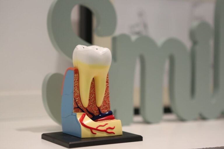
Above the upper premolars and molars, the sinus floor can be as thin as a few millimetres. In some people, the roots of these teeth actually protrude into the sinus cavity. This close relationship explains why dental work in the upper jaw sometimes affects the sinus.
How extractions can affect the sinus
During a routine extraction, the dentist or oral surgeon lifts the tooth away from the surrounding bone. If the bone is thin or the tooth is impacted, the instrument can accidentally push through the sinus floor, creating a perforation. The size of the opening can range from a pin‑hole to a larger gap that allows air and fluids to move between the mouth and the sinus.
Most small perforations seal themselves within a few days, but larger ones may stay open, allowing bacteria from the mouth to enter the sinus. This can lead to infection, chronic sinusitis, or a persistent foul taste.
Recognizing Early Warning Signs
Common symptoms
After an upper tooth extraction, some discomfort is normal. However, certain signs suggest a sinus involvement:
- Clear, watery nasal discharge that increases when you bend over.
- Yellow or green discharge that may have a foul smell.
- A salty or metallic taste in the mouth, especially after drinking.
- Feeling of pressure or fullness in the cheek or around the eye.
- Pain that radiates from the extraction site up into the sinus area.
- Unexplained nosebleeds or occasional coughing.
When symptoms may indicate a perforation
If any of the above symptoms appear within the first few days after extraction and do not improve with routine post‑operative care, a sinus perforation should be considered. Persistent drainage that does not stop after a week, worsening facial pain, or a sudden change in the colour of the discharge are red flags that merit a dental or medical visit.
It is also important to note that some patients may experience a “dry socket” – a painful condition where the blood clot dissolves. While dry socket shares some pain features, it does not usually cause nasal discharge. Distinguishing between the two helps guide appropriate treatment.

Detailed Symptom Breakdown
The following table summarises the most frequently reported symptoms, their typical onset, and what they may indicate about the underlying issue.
| Symptom | Typical Onset | Possible Meaning |
|---|---|---|
| Clear nasal discharge | 1‑3 days post‑extraction | Minor sinus communication; often resolves quickly |
| Thick, coloured discharge | 4‑7 days post‑extraction | Possible infection or larger perforation |
| Foul taste or smell | 2‑5 days post‑extraction | Oral‑sinus communication allowing food particles into sinus |
| Facial pressure or headache | Within first week | Sinus inflammation or blockage |
| Persistent pain at extraction site | Beyond 5‑7 days | Could be dry socket, infection, or perforation‑related irritation |
While the table provides a quick reference, individual experiences vary. If you notice any combination of these signs, especially drainage that changes in colour or consistency, reach out to a health professional promptly.
How Doctors Diagnose a Sinus Perforation
Physical examination
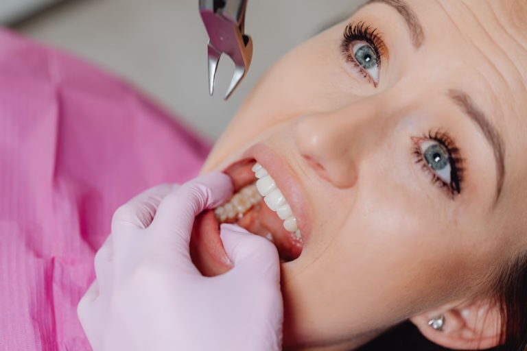
The dentist or oral surgeon will first inspect the extraction site for any visible opening. They may use a small mirror and light to look for fluid leakage into the mouth. Palpating the cheek and asking you to blow your nose can also reveal a communication between the mouth and sinus.
In some cases, the clinician will gently press on the sinus area while you swallow. A sudden rush of fluid into the mouth or a change in pressure sensation can confirm a perforation.
Imaging options
Imaging is the most reliable way to visualise the perforation. Common modalities include:
- Panoramic X‑ray (OPG): Provides a broad view of the jaws and can show obvious bone loss.
- Cone‑beam computed tomography (CBCT): Offers three‑dimensional detail of the sinus floor and the exact size of the opening.
- Standard CT scan: Used when infection is suspected, as it also shows fluid collections.
These images help the clinician decide whether the perforation will heal on its own or needs surgical closure. For more information on imaging techniques, you can visit the MedlinePlus health information portal.

Managing Symptoms at Home
While waiting for a professional appointment, several self‑care steps can reduce discomfort and lower the risk of infection.
- Keep your head elevated when lying down to minimise sinus pressure.
- Avoid blowing your nose forcefully for at least 48 hours after extraction.
- Rinse gently with a warm saline solution using a syringe (no suction) to keep the area clean.
- Stay hydrated; water helps thin mucus and promotes drainage.
- Use over‑the‑counter pain relievers such as ibuprofen, unless contraindicated, to control pain and inflammation.
- Apply a warm compress to the cheek for 10‑15 minutes, three times a day, to soothe pressure.
Professional Treatment Options
Small perforations – conservative care
For perforations smaller than 2 mm, many clinicians recommend a watch‑and‑wait approach. The sinus membrane often seals itself within a week or two. During this period, the dentist may prescribe a short course of antibiotics to prevent bacterial invasion and may advise nasal decongestants to keep the sinus drainage pathways open.
Follow‑up appointments are essential to confirm that the opening has closed. A repeat CBCT scan after two weeks can demonstrate healing.
Larger perforations – surgical repair
When the opening exceeds 2‑3 mm or when symptoms persist despite conservative care, surgical intervention is considered. The most common techniques include:
- Buccal flap closure: Tissue from the cheek is rotated to cover the perforation.
- Sinus membrane graft: A thin layer of collagen or autologous tissue is placed over the defect.
- Endoscopic sinus surgery: Performed by an ENT specialist to repair the sinus floor and address any concurrent sinusitis.
These procedures are usually done under local or general anaesthesia and have a high success rate. Post‑operative care mirrors the home‑care steps listed earlier, with added instructions on keeping the surgical site clean.
Preventing Future Problems
Even though sinus perforation is relatively uncommon, certain precautions can lower the risk during future dental work:
- Ask your dentist to review any prior imaging that shows the proximity of your tooth roots to the sinus.
- Inform the clinician if you have a history of chronic sinusitis or recent upper‑respiratory infections.
- Consider a pre‑operative CBCT scan for upper molar extractions, especially if the teeth are impacted.
- Follow post‑operative instructions meticulously, avoiding activities that increase sinus pressure such as heavy lifting or forceful nose blowing.
- Maintain good oral hygiene to reduce bacterial load that could migrate into the sinus.
Frequently Asked Questions
Can a sinus perforation heal without surgery?
Yes, many small perforations seal on their own within 7‑10 days, especially when proper oral hygiene and gentle nasal care are followed. Monitoring symptoms and attending follow‑up appointments are key.
Is it dangerous to have a sinus perforation?
While most cases are not life‑threatening, an untreated perforation can lead to chronic sinus infection, persistent foul taste, or even spread of infection to surrounding bone (osteomyelitis). Prompt evaluation reduces these risks.
Will I need antibiotics?
Antibiotics are often prescribed when there is evidence of bacterial infection or when the perforation is larger. Your dentist will decide based on clinical findings and imaging.
How long before I can eat normally?
Soft foods are recommended for the first 3‑4 days. As the site heals and any discharge subsides, you can gradually reintroduce regular foods. Avoid very hot, spicy, or acidic items that could irritate the area.
The Bottom Line
Sinus perforation after tooth extraction symptoms are a specific set of nasal and oral signs that signal a communication between the mouth and the maxillary sinus. Early recognition, appropriate imaging, and timely treatment, whether conservative or surgical, help prevent complications and ensure a smooth recovery. If you notice persistent discharge, foul taste, or facial pressure after an upper tooth extraction, contact a dental or medical professional right away.
Was this page helpful?
Thanks for your feedback! What could we improve?
Thank you! Your feedback helps us improve our articles.
Medically reviewed by
Dr. Bernard ReynoldsPhysician with experience in patient care and preventive medicine, focused on early detection, disease prevention and long-term wellness.
True Impact Health articles are written by our editorial team and based on reliable sources such as public health agencies, clinical guidelines and peer-reviewed research. This article has been checked by a doctor on our review panel. Read our editorial process.
This content is for general information and is not a substitute for professional medical advice, diagnosis or treatment.