Introduction
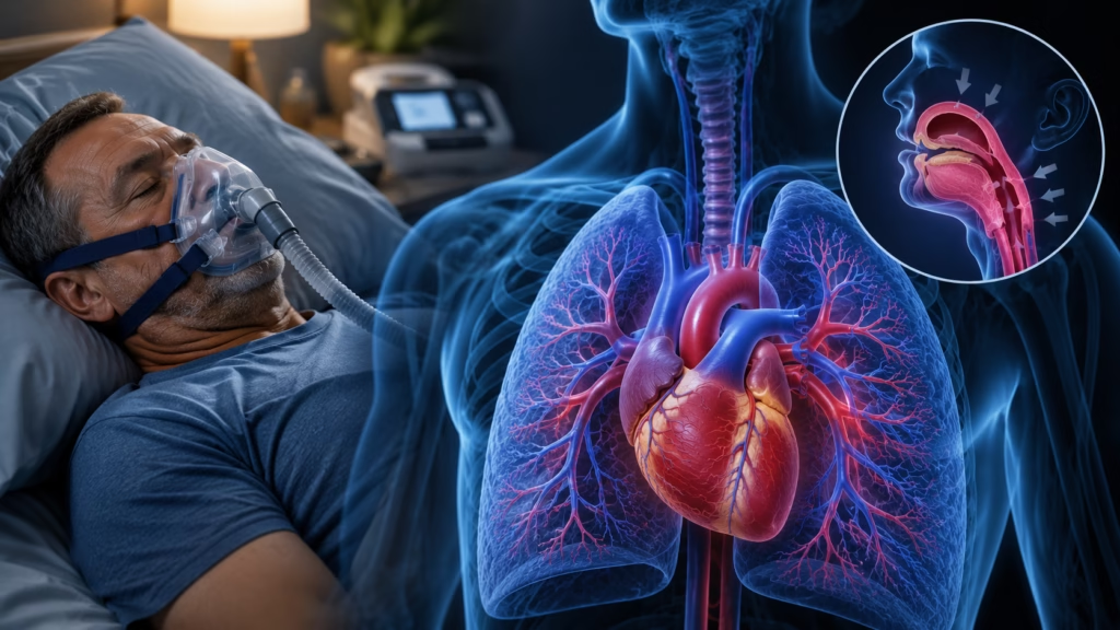
Pulmonary hypertension (PH) is a serious and progressive cardiovascular disorder that affects the blood vessels connecting the heart and lungs. As pressure within these vessels rises, the right side of the heart is forced to work harder to pump blood through the lungs. Over time, this increased workload can weaken the heart, reduce exercise capacity, and significantly affect a person’s quality of life.
One of the most important measurements used during the evaluation of pulmonary hypertension is the Right Ventricular Systolic Pressure (RVSP). This value, typically estimated through echocardiography, provides valuable information about the pressure within the pulmonary circulation and the amount of stress being placed on the right ventricle. Although RVSP is not a direct measurement of pulmonary artery pressure, it serves as a useful non-invasive marker that helps physicians identify possible pulmonary hypertension, assess disease severity, and monitor treatment response.
Understanding RVSP can help patients better interpret echocardiogram results and appreciate how physicians evaluate pulmonary hypertension. This guide explains what RVSP is, how it is measured, what normal and abnormal values mean, and how this important parameter influences diagnosis and treatment decisions.
Understanding Pulmonary Hypertension
Pulmonary hypertension is a condition characterized by abnormally high blood pressure within the pulmonary arteries, the blood vessels responsible for carrying blood from the right side of the heart to the lungs. Unlike systemic blood pressure, which is measured in the arteries throughout the body, pulmonary artery pressure specifically reflects the pressure within the lung circulation.
Under normal circumstances, blood flows easily through the pulmonary arteries, allowing oxygen exchange to occur efficiently within the lungs. However, in pulmonary hypertension, these arteries may become narrowed, thickened, stiffened, or obstructed. As resistance within the pulmonary circulation increases, the right ventricle must generate greater force to maintain adequate blood flow.
Initially, the right ventricle adapts by becoming thicker and stronger. However, prolonged pressure overload eventually causes the chamber to enlarge and weaken. If left untreated, this process can progress to right-sided heart failure, which is one of the most serious complications of pulmonary hypertension.
Related Posts:


Pulmonary hypertension is classified into five major groups according to the World Health Organization. These include pulmonary arterial hypertension, pulmonary hypertension caused by left-sided heart disease, pulmonary hypertension associated with lung diseases, chronic thromboembolic pulmonary hypertension, and pulmonary hypertension resulting from multifactorial or unclear mechanisms. Although the causes vary, all forms ultimately increase the workload of the right ventricle.
Patients often experience symptoms such as shortness of breath during physical activity, fatigue, dizziness, chest discomfort, swelling of the legs, and reduced exercise tolerance. Because these symptoms can develop gradually and mimic other conditions, pulmonary hypertension is frequently diagnosed late in the disease course.
What Is Right Ventricular Systolic Pressure (RVSP)?
Right Ventricular Systolic Pressure, commonly abbreviated as RVSP, refers to the maximum pressure generated by the right ventricle during contraction. During each heartbeat, the right ventricle pumps blood into the pulmonary arteries, and RVSP reflects the force required to accomplish this task.
In individuals without significant obstruction of the pulmonary valve, RVSP closely approximates Pulmonary Artery Systolic Pressure (PASP). For this reason, RVSP is widely used as a non-invasive estimate of pulmonary artery pressure during echocardiographic evaluation.
RVSP provides important information because it reflects how hard the right side of the heart must work. As pulmonary artery pressure rises, the right ventricle must generate higher pressures to overcome increased resistance within the lungs. Consequently, elevated RVSP values often indicate pulmonary hypertension or other conditions that place excessive strain on the right side of the heart.
For physicians, RVSP serves as an important screening tool that can identify patients who require further investigation. For patients, RVSP offers insight into disease severity and helps track changes over time.
How RVSP Is Measured
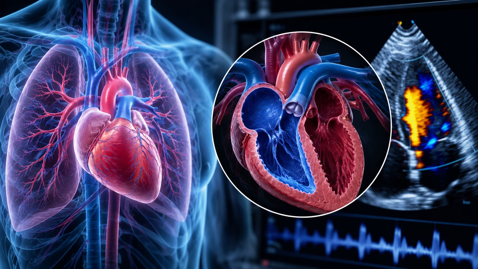
The most common method for estimating RVSP is Doppler echocardiography, a non-invasive ultrasound examination of the heart. Echocardiography allows physicians to visualize heart structures, assess cardiac function, and estimate blood flow velocities throughout the heart.
The calculation of RVSP relies primarily on the presence of tricuspid regurgitation, which occurs when a small amount of blood leaks backward through the tricuspid valve during ventricular contraction. This leakage creates a measurable jet of blood that can be detected using Doppler ultrasound.
By measuring the velocity of this regurgitant jet, clinicians can estimate the pressure difference between the right ventricle and the right atrium using the modified Bernoulli equation:
\Delta P = 4V^2
In this equation, ΔP represents the pressure gradient across the tricuspid valve, while V represents the velocity of the tricuspid regurgitation jet.
After calculating the pressure gradient, physicians estimate right atrial pressure based on the size and respiratory movement of the inferior vena cava, a large vein that returns blood to the heart. The estimated right atrial pressure is then added to the tricuspid valve gradient.
The final equation for RVSP is:
RVSP = TR\ Gradient + RAP
For example, if the tricuspid regurgitation velocity is 3.0 meters per second, the pressure gradient would equal 36 mmHg. If the estimated right atrial pressure is 10 mmHg, the calculated RVSP would be approximately 46 mmHg.
This non-invasive approach provides valuable information without requiring invasive procedures.
Normal RVSP Values
In healthy adults, RVSP generally falls below 35 mmHg. Values within this range usually indicate normal pulmonary artery pressures and normal right ventricular workload.
However, RVSP values must always be interpreted within the broader clinical context. Factors such as age, body size, obesity, lung disease, and technical limitations during echocardiography can influence measurements. Therefore, a mildly elevated RVSP does not automatically confirm pulmonary hypertension.
Many cardiologists and pulmonologists consider RVSP values exceeding 35 to 40 mmHg as potentially abnormal and deserving of further evaluation, especially when accompanied by symptoms or additional echocardiographic abnormalities.
What Elevated RVSP Indicates
An elevated RVSP suggests increased pressure within the pulmonary circulation and increased strain on the right ventricle.
Mild elevations typically range from approximately 35 to 50 mmHg. At this stage, patients may have minimal symptoms or experience only mild exertional shortness of breath. Although the condition may still be relatively early, further investigation is generally warranted.
Moderate elevations usually range from 50 to 70 mmHg. Patients often begin experiencing more noticeable limitations in exercise tolerance, fatigue, and breathlessness. Structural changes within the right ventricle may become evident during imaging studies.
Severe elevations generally exceed 70 mmHg and often indicate advanced pulmonary hypertension. At this stage, the right ventricle may show significant enlargement and dysfunction. Symptoms can become severe and may include shortness of breath at rest, fluid retention, dizziness, and signs of right-sided heart failure.
Higher RVSP values have been associated with increased hospitalization rates, worsening symptoms, and greater mortality risk. For this reason, identifying elevated RVSP early is critical for timely intervention.
The Relationship Between RVSP and Right Heart Function
One of the most important aspects of RVSP assessment is its relationship to right ventricular function.
As pulmonary artery pressure increases, the right ventricle must generate increasingly higher pressures to maintain blood flow. Initially, the heart compensates by thickening its muscular walls, a process known as hypertrophy. While this adaptation may temporarily preserve cardiac output, chronic pressure overload eventually leads to ventricular enlargement and weakening.
Elevated RVSP often serves as an early warning sign that the right ventricle is under stress. Monitoring RVSP over time allows physicians to detect disease progression before significant heart failure develops.
In many patients, worsening RVSP correlates with declining exercise capacity, increasing symptom burden, and reduced quality of life.
How RVSP Fits Into the Diagnostic Process
Although RVSP is an important measurement, it is only one component of a comprehensive pulmonary hypertension evaluation.
A complete assessment typically begins with a detailed medical history and physical examination. Physicians evaluate symptoms, risk factors, family history, and signs of right-sided heart strain.
Blood tests such as BNP or NT-proBNP may be ordered to assess cardiac stress and heart failure severity. Imaging studies including chest X-rays and CT scans help identify structural lung or heart abnormalities.
Echocardiography remains the most important initial imaging test because it estimates RVSP, evaluates right ventricular size and function, and identifies other cardiac abnormalities that may contribute to pulmonary hypertension.
However, the definitive diagnosis of pulmonary hypertension requires right heart catheterization. This invasive procedure directly measures pulmonary artery pressures and provides precise hemodynamic information. Right heart catheterization remains the gold standard because echocardiographic RVSP estimates can occasionally overestimate or underestimate actual pressures.
How RVSP Influences Treatment Decisions
RVSP plays a significant role in guiding treatment strategies.
Patients with mild elevations may initially undergo observation, risk-factor modification, and treatment of underlying conditions. Lifestyle adjustments, oxygen therapy, and management of sleep apnea or lung disease may be sufficient in some cases.
More severe elevations often require targeted pulmonary hypertension therapies. These may include endothelin receptor antagonists, phosphodiesterase-5 inhibitors, prostacyclin analogues, or soluble guanylate cyclase stimulators. These medications aim to reduce pulmonary vascular resistance and lower pressure within the pulmonary circulation.
As treatment progresses, repeat echocardiograms help monitor RVSP trends. A declining RVSP may indicate successful therapy, whereas increasing values may signal disease progression or inadequate response to treatment.
For patients with advanced disease, RVSP monitoring helps determine when more aggressive interventions such as lung transplantation or advanced surgical procedures should be considered.
Living With Pulmonary Hypertension and Monitoring RVSP
Managing pulmonary hypertension is a long-term process that requires ongoing monitoring and collaboration between patients and healthcare providers.
Regular echocardiograms are often performed to evaluate RVSP and assess right ventricular function. These studies provide valuable information about disease stability and treatment effectiveness.
Patients may also undergo six-minute walk tests, cardiopulmonary exercise testing, and laboratory evaluations to monitor overall functional status.
Lifestyle modifications play an important role in disease management. Maintaining a healthy weight, limiting sodium intake, adhering to prescribed medications, staying physically active within recommended limits, and avoiding smoking can all contribute to improved outcomes.
Because pulmonary hypertension can affect emotional well-being, psychological support and patient education are also important components of comprehensive care.
Conclusion
Right Ventricular Systolic Pressure (RVSP) is one of the most valuable non-invasive measurements used in the evaluation of pulmonary hypertension. By estimating the pressure generated by the right ventricle during contraction, RVSP provides important insight into pulmonary artery pressures and the overall burden placed on the right side of the heart.
Although RVSP alone cannot definitively diagnose pulmonary hypertension, elevated values often serve as an early warning sign that further evaluation is needed. Through echocardiography, physicians can use RVSP to screen for pulmonary hypertension, assess disease severity, guide treatment decisions, and monitor response to therapy over time.
For patients living with pulmonary hypertension, understanding RVSP can provide a clearer picture of disease progression and treatment goals. Regular monitoring, timely intervention, and close communication with healthcare providers remain essential for achieving the best possible outcomes and preserving quality of life.
Disclaimer
This article is for educational purposes only and does not replace professional medical advice, diagnosis, or treatment. Seizures, severe headache, confusion, visual changes, or blood pressure readings in the hypertensive crisis range require urgent medical evaluation.