
















Spinal stenosis
Spinal stenosis is a common cause of back pain and leg weakness; learn symptoms, diagnosis, and treatment options in this clear, evidence‑based guide.
By True Impact Health Editorial Team

Medical graduate of Girne American University with a focus on patient-centred care, clinical assessment and preventive health.
Reviewed this article on November 5, 2026.
View profile How we reviewUpdated on November 4, 2026 9 min read

Table of Contents
Key Takeaways
- Spinal stenosis often results from age‑related wear and tear
- Typical symptoms include pain, numbness, and balance problems
- Diagnosis relies on physical exam and imaging such as MRI
- Treatment ranges from physical therapy to surgery, tailored to severity
Spinal stenosis is a condition where the spaces within the spine narrow, putting pressure on the spinal cord or nerves. This narrowing can cause pain, tingling, or weakness in the back, legs, or arms, depending on where it occurs. Understanding what spinal stenosis is, why it happens, and how it can be managed helps anyone facing these symptoms feel more in control.
The condition is most common in older adults, but it can affect younger people who have injuries or congenital spine issues. While the word sounds technical, the experience is often familiar: a dull ache that worsens with walking, relief when sitting, and occasional sharp shocks down the limbs. This guide explains the condition in plain language, using up‑to‑date medical guidance.
By the end of this article you will know the typical causes, warning signs, how doctors confirm the diagnosis, and what treatment paths are available, from simple home measures to surgical options.
What Is Spinal Stenosis?
Definition
Spinal stenosis describes a narrowing of the central canal, the lateral recesses, or the intervertebral foramina, the openings through which nerves exit the spine. When these passages shrink, they can compress the spinal cord itself (central stenosis) or the nerve roots (foraminal stenosis). The compression interferes with normal nerve signaling, leading to pain, numbness, or weakness.
Types of Spinal Stenosis
The spine is divided into three regions. Cervical stenosis occurs in the neck and can affect the arms and hands. Thoracic stenosis is rare and involves the mid‑back, sometimes causing chest or abdominal symptoms. Lumbar stenosis, the most frequent form, affects the lower back and typically produces leg pain, known as neurogenic claudication.
Each region shares the same basic mechanism, space loss, but the symptoms differ because of the nerves that travel through each area.
Causes and Risk Factors
Age‑Related Changes
As we age, the discs between vertebrae lose water content and become thinner. The facet joints, which help the spine move, develop arthritis that can produce bone spurs. These spurs, together with thickened ligaments, encroach on the spinal canal. The cumulative effect of these changes is the most common cause of spinal stenosis in people over 50.
Other Contributing Factors
In addition to natural wear, several conditions increase risk. Congenital spinal stenosis means the canal was narrow from birth. Past spine injuries, such as fractures or dislocations, can alter the anatomy and trigger stenosis later. Certain genetic disorders, like achondroplasia, also predispose individuals to a smaller spinal canal.
Lifestyle factors play a role as well. Smoking reduces blood flow to spinal tissues, accelerating degeneration. Obesity adds mechanical load to the spine, hastening joint wear. Maintaining a healthy weight and avoiding tobacco can therefore lower long‑term risk.

Symptoms to Watch For
Symptoms vary by location but share a pattern of activity‑related discomfort that eases with rest. Common signs include:
- Back pain that worsens when standing or walking
- Numbness, tingling, or “pins‑and‑needles” in the legs or arms
- Weakness that makes it hard to climb stairs or lift objects
- Balance problems, especially on uneven ground
- Relief of symptoms when leaning forward or sitting
In cervical stenosis, hand clumsiness, dropping objects, or difficulty with fine motor tasks may appear. Thoracic stenosis can cause a band‑like sensation around the chest or abdomen.
Because these signs overlap with other back problems, a professional evaluation is essential to confirm spinal stenosis.
How Is It Diagnosed?
Physical Examination
A clinician begins with a detailed history and a physical exam. They assess gait, reflexes, muscle strength, and sensation. Specific maneuvers, such as the “straight leg raise” or “Spurling’s test,” help differentiate nerve compression from other causes.
Imaging Tests
Imaging provides a visual map of the spine’s anatomy. The most common studies include:
| Test | What It Shows | Typical Use |
|---|---|---|
| MRI | Detailed soft‑tissue images | Gold standard for spinal canal size |
| CT scan | Bone detail and canal dimensions | When MRI unavailable |
| X‑ray | Bone alignment, arthritis | Initial screening |
MRI is preferred because it visualises both bone and soft tissue, revealing disc bulges, ligament thickening, and nerve compression without radiation. CT can be useful if MRI is contraindicated, such as in patients with certain implants.
Blood tests are not diagnostic for stenosis but may be ordered to rule out inflammatory arthritis or infection.
Treatment Options
Conservative Management
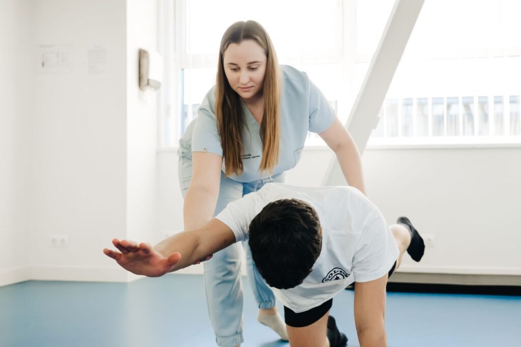
Most people start with non‑surgical approaches. Physical therapy focuses on strengthening core muscles, improving flexibility, and teaching posture techniques that reduce pressure on the spine. A therapist may also use modalities like heat, gentle traction, or electrical stimulation to ease pain.
Medications can help control symptoms. Over‑the‑counter pain relievers such as acetaminophen or non‑steroidal anti‑inflammatory drugs (NSAIDs) reduce inflammation and discomfort. In some cases, doctors prescribe muscle relaxants or low‑dose antidepressants that have pain‑modulating properties.
Activity modification is another key element. Walking, swimming, or cycling are low‑impact exercises that maintain fitness without aggravating the spine. Patients are often advised to avoid prolonged standing or heavy lifting.
Interventional Procedures
When symptoms persist despite therapy, injections may be considered. Epidural steroid injections deliver anti‑inflammatory medication directly around the compressed nerves, offering temporary relief that can last weeks to months.
Facet joint injections target painful arthritic joints. These procedures are performed under imaging guidance to ensure accurate placement.
While injections do not cure stenosis, they can reduce pain enough to allow continued participation in physical therapy, which may improve long‑term outcomes.
Surgical Options
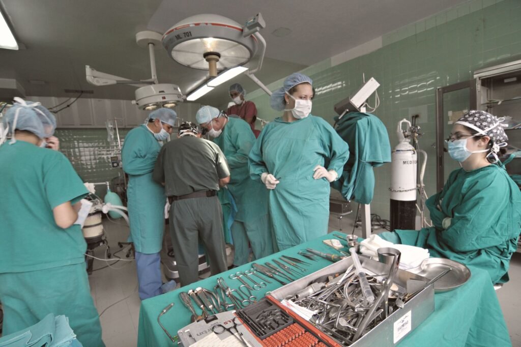
Surgery is reserved for moderate to severe cases where quality of life is significantly impacted or neurological deficits worsen. The most common operation is a decompressive laminectomy, where the surgeon removes part of the vertebral bone (lamina) to enlarge the canal.
In some patients, spinal fusion is added to stabilize the segment after bone removal. Minimally invasive techniques use smaller incisions and specialized instruments, resulting in shorter hospital stays and quicker recovery.
Outcomes are generally good, with many patients reporting reduced pain and improved walking distance. However, surgery carries risks such as infection, blood loss, and the possibility of needing additional procedures later.
Lifestyle Strategies for Managing Symptoms
Beyond formal treatment, everyday habits can make a big difference. Here are practical steps anyone can adopt:
- Maintain a healthy weight. Extra pounds increase load on the lumbar spine, accelerating wear.
- Practice good posture. Use lumbar support when sitting and keep screens at eye level to avoid forward head posture.
- Incorporate core‑strengthening exercises. Planks, bridges, and gentle Pilates improve spinal stability.
- Stay hydrated. Healthy discs rely on water to maintain height and cushioning.
- Quit smoking. Smoking impairs blood flow to spinal tissues, worsening degeneration.
Heat or cold packs applied to the painful area can provide temporary relief. A warm shower or heating pad relaxes tight muscles, while an ice pack reduces acute inflammation after activity.
Finally, consider ergonomic adjustments at work. A standing desk, footrest, or chair with adjustable lumbar support can reduce prolonged strain.
When to Seek Medical Help
If you notice any of the following, contact a healthcare professional promptly:
- Sudden, severe back pain after a fall or injury.
- Progressive weakness that makes it difficult to lift your foot or grip objects.
- Loss of bladder or bowel control, which may signal a medical emergency.
- Pain that does not improve with rest, over‑the‑counter medication, or activity modification after a few weeks.
Early evaluation allows for timely intervention, which can prevent irreversible nerve damage and preserve function.
Advanced Imaging and Assessment
MRI versus CT Scan
Magnetic resonance imaging provides detailed views of soft tissues, nerve roots, and the spinal canal, making it the preferred first‑line study for most patients. Computed tomography excels at visualizing bone anatomy and can clarify bony stenosis when MRI is contraindicated. Selecting the appropriate modality depends on the clinical question, patient safety, and availability of equipment.
Dynamic Flexion‑Extension X‑rays
These studies capture the spine in motion, revealing instability that static images may miss. They are particularly useful for patients with a history of trauma or those who exhibit worsening symptoms with certain movements. Radiologists compare the vertebral alignment in flexion and extension to assess for abnormal translation.
- Identify occult instability
- Guide surgical planning
- Assess post‑operative alignment
Targeted Physical Therapy Approaches
Core Stabilization Exercises
Strengthening the deep abdominal and lumbar muscles helps maintain spinal alignment and reduces load on narrowed canals. Common drills include pelvic tilts, bird‑dogs, and modified planks performed with controlled breathing. Consistency, rather than intensity, yields the best long‑term outcomes.
Neurodynamic Mobilization
These techniques gently glide nerve roots to improve their mobility and reduce irritation. Therapists use specific limb movements combined with spinal positioning to create tension‑release cycles. When performed correctly, patients often notice decreased radicular pain after several sessions.
Aquatic Therapy
Water buoyancy lessens axial loading, allowing patients to practice range‑of‑motion exercises with less discomfort. Walking in a pool, water‑based leg lifts, and resistance band work are typical components. The warm environment also promotes muscle relaxation.
- Reduced joint stress
- Improved cardiovascular fitness
- Enhanced proprioception
Emerging Surgical Techniques and Recovery
Minimally Invasive Decompression
Through tubular retractors and endoscopic cameras, surgeons can remove offending bone and ligament without large incisions. Reported benefits include less blood loss, shorter hospital stays, and quicker return to daily activities. Patient selection remains critical; the technique is most effective for focal stenosis.
Interspinous Process Devices
These implants act as spacers that distract the vertebrae, enlarging the canal while preserving motion. They are typically placed percutaneously under fluoroscopic guidance. Long‑term data suggest modest symptom relief for patients who are not candidates for fusion.
Post‑operative Rehabilitation Timeline
Early mobilization begins within 24‑48 hours, focusing on gentle walking and breathing exercises. By week two, patients progress to low‑impact strengthening and flexibility work. Full functional recovery often occurs between six and twelve weeks, depending on the extent of the procedure.
- Week 1: Light ambulation, diaphragmatic breathing
- Weeks 2‑4: Core activation, gentle stretching
- Weeks 5‑12: Progressive resistance training, functional tasks
Frequently Asked Questions
Can spinal stenosis be prevented?
While the natural aging process cannot be stopped, risk can be lowered by staying active, maintaining a healthy weight, avoiding smoking, and managing chronic conditions such as diabetes that affect tissue health.
Is surgery always required?
No. Most people manage symptoms with physical therapy, medication, and lifestyle changes. Surgery is considered when conservative care fails or neurological deficits worsen.
How long does recovery take after surgery?
Recovery varies. Many patients resume light activities within 4‑6 weeks, while full return to work may take 3‑6 months, depending on the procedure and individual health.
Will spinal stenosis ever fully go away?
Spinal stenosis is a chronic condition. Treatment aims to reduce symptoms and improve function, not necessarily to eliminate the narrowing. Ongoing self‑care helps keep symptoms at a manageable level.
The Bottom Line
Spinal stenosis is a common, age‑related narrowing of the spinal canal that can cause pain, numbness, and functional limitations. Diagnosis relies on a careful exam and imaging, most often MRI. Treatment starts with conservative measures, exercise, medication, and lifestyle tweaks, and progresses to injections or surgery when needed. Early recognition and a proactive approach empower individuals to maintain mobility and quality of life.MedlinePlus
Was this page helpful?
Thanks for your feedback! What could we improve?
Thank you! Your feedback helps us improve our articles.
Medically reviewed by
Dr. Oluwabusola Stella OkeMedical graduate of Girne American University with a focus on patient-centred care, clinical assessment and preventive health.
True Impact Health articles are written by our editorial team and based on reliable sources such as public health agencies, clinical guidelines and peer-reviewed research. This article has been checked by a doctor on our review panel. Read our editorial process.
This content is for general information and is not a substitute for professional medical advice, diagnosis or treatment.