









Bursitis
Bursitis explained: symptoms, causes, diagnosis, and treatment options to help you manage joint pain and get back to daily activities.
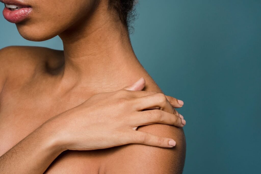
Table of Contents
Key Takeaways
- Bursitis is inflammation of fluid‑filled sacs that cushion joints.
- Common sites include shoulder, elbow, hip, and knee.
- Treatment starts with rest, ice, and over‑the‑counter pain relievers.
- Seek medical evaluation if pain persists or you notice swelling or fever.
Bursitis is a condition that many people encounter when the small, fluid‑filled sacs called bursae become inflamed. These bursae act as cushions between bones, tendons, and muscles, reducing friction during movement. When they swell, everyday activities such as lifting, walking, or even sleeping can become painful.
Understanding bursitis helps you recognize early signs, seek appropriate care, and adopt strategies that speed recovery. This guide covers everything from causes and symptoms to diagnosis, treatment, and self‑care tips, all written in plain language for readers without a medical background.
What Is Bursitis?
Definition
Bursitis refers to inflammation of a bursa, a tiny sac filled with lubricating fluid. The bursa’s job is to allow smooth gliding of tissues over each other. When irritation, overuse, or infection occurs, the sac fills with excess fluid, causing swelling, warmth, and pain.
Causes
The most frequent trigger is repetitive motion or prolonged pressure on a joint. Examples include frequent overhead lifting, leaning on elbows for long periods, or repetitive kneeling. Direct injury, such as a fall onto a joint, can also inflame the bursa. In some cases, bacteria enter the bursa through a skin cut, leading to septic bursitis, which requires prompt medical attention.
Who Is at Risk?
Anyone can develop bursitis, but certain groups are more prone. Athletes who perform repetitive motions, tennis players, swimmers, and weightlifters, often experience shoulder or elbow bursitis. Workers who kneel or squat for hours, such as gardeners or carpet installers, may develop knee or hip bursitis. Age‑related wear and tear, rheumatoid arthritis, and gout also increase susceptibility.
Common Locations and Symptoms
Shoulder (Subacromial Bursitis)
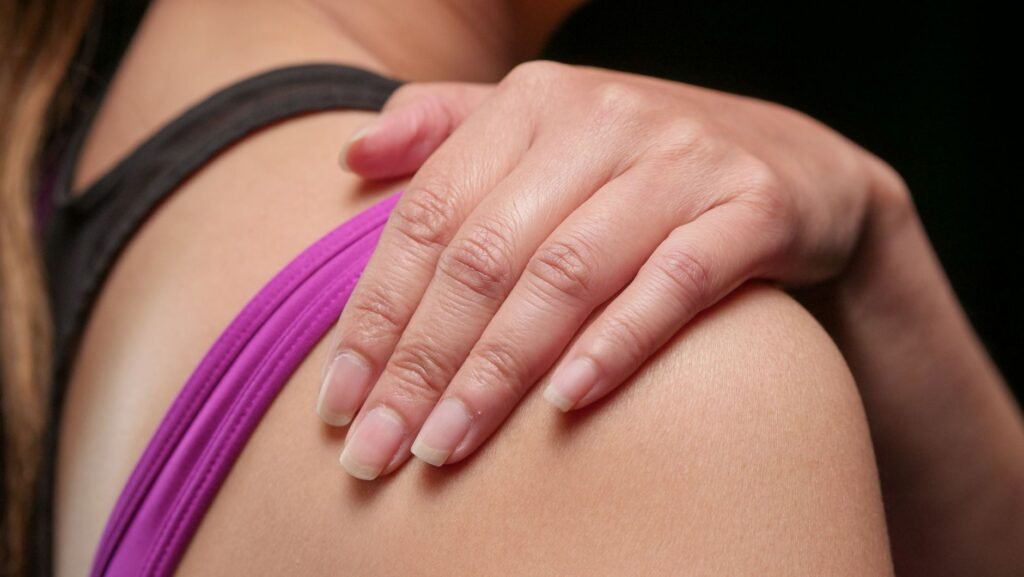
Shoulder bursitis typically feels like a deep ache on the top of the arm, sometimes spreading down the side. Raising the arm overhead may be painful, and a noticeable lump can develop over the acromion.
Elbow (Olecranon Bursitis)
Also called “student’s elbow,” this form produces a soft, swollen bump on the tip of the elbow. The area may feel warm, and pressing on it often elicits tenderness.
Hip (Trochanteric Bursitis)
Hip bursitis causes pain on the outer thigh and hip, especially when walking or lying on the affected side. The pain may worsen at night, making it hard to fall asleep.
Knee (Prepatellar Bursitis)
Often called “housemaid’s knee,” this type presents as a visible swelling in front of the kneecap. It can be tender to touch and may feel stiff after prolonged kneeling.
| Joint | Typical Symptoms | Common Triggers |
|---|---|---|
| Shoulder | Aching, limited overhead movement, lump | Overhead sports, heavy lifting |
| Elbow | Swelling tip, warmth, tenderness | Resting elbows on hard surfaces |
| Hip | Outer thigh pain, night discomfort | Running, prolonged standing |
| Knee | Front‑kneecap swelling, stiffness | Kneeling, crawling |
How Bursitis Is Diagnosed
Physical Examination
A clinician will first ask about your symptoms, activity history, and any recent injuries. They will gently palpate the affected area to feel for swelling, warmth, and tenderness. Specific movements are tested to see which actions worsen the pain, helping narrow down the exact bursa involved.
Imaging Tests
While many cases are diagnosed clinically, imaging can confirm the diagnosis or rule out other problems. Ultrasound is useful for visualizing fluid in the bursa and guiding needle aspiration if needed. MRI provides detailed images of soft tissues and can differentiate bursitis from tendon tears or arthritis.
Laboratory Tests
If infection is suspected, a doctor may aspirate fluid from the bursa using a thin needle. The fluid is examined under a microscope and cultured to identify bacteria. Blood tests may also be ordered to look for signs of systemic inflammation.
Treatment Options
Rest and Activity Modification
Giving the inflamed bursa time to heal is the cornerstone of treatment. Avoid activities that aggravate the joint for several days to weeks. Modifying workstations, using padded supports, or switching to low‑impact exercises can reduce stress on the affected area.
Ice, Heat, and Compression
Applying ice packs for 15‑20 minutes several times a day in the first 48‑72 hours helps limit swelling. After the acute phase, gentle heat can relax surrounding muscles. Elastic bandages or compression sleeves provide mild support without restricting circulation.
Medications
Over‑the‑counter non‑steroidal anti‑inflammatory drugs (NSAIDs) such as ibuprofen or naproxen can relieve pain and reduce inflammation. Topical NSAID gels are an alternative for those who cannot take oral medication. Always follow label directions and consult a pharmacist if you have health conditions.
Physical Therapy
Targeted stretching and strengthening exercises improve joint mechanics and prevent recurrence. A therapist may teach you rotator‑cuff strengthening for shoulder bursitis or hip‑abductor exercises for trochanteric bursitis. Proper technique is essential to avoid further irritation.
Injections and Surgery
If symptoms persist despite conservative care, a healthcare provider might inject corticosteroids directly into the bursa to rapidly reduce inflammation. In rare, chronic cases where the bursa is severely damaged, surgical removal (bursectomy) may be considered.
Home Care and Self‑Management
- Apply ice for the first few days, then switch to heat.
- Take NSAIDs with food to protect your stomach.
- Use a supportive pillow or cushion when sleeping on the affected side.
- Perform gentle range‑of‑motion exercises several times a day.
- Maintain a healthy weight to lessen joint load.
- Stay hydrated; adequate fluid supports joint health.
Consistent self‑care often speeds recovery and reduces the chance of flare‑ups. However, if pain worsens, swelling spreads, or you develop fever, seek medical attention promptly.
When to Seek Professional Help
If pain does not improve within a week of home treatment, or if you notice any of the following, contact a healthcare professional:
- Increasing swelling or warmth over the joint.
- Fever, chills, or red streaks spreading from the area.
- Severe pain that limits basic activities such as dressing or walking.
- Loss of strength or numbness in the limb.
Early evaluation can identify septic bursitis, which requires antibiotics, or other conditions that may mimic bursitis, such as arthritis or tendonitis.
Risk Factors and Prevention
Modifiable Lifestyle Factors
Weight gain increases pressure on weight‑bearing joints and can irritate the subacromial and trochanteric bursae. Repetitive activities such as prolonged kneeling or overhead lifting create micro‑trauma that predisposes the bursa to inflammation. Smoking reduces tissue oxygenation and impairs healing, making bursitis more likely after a minor injury. Regular aerobic exercise and maintaining a healthy body mass index help keep the surrounding muscles supportive and reduce stress on bursae.
Occupational and Sports‑related Risks
Jobs that require frequent crouching, pushing, or pulling place the shoulder, elbow, and hip bursae under constant strain. Athletes in tennis, volleyball, or rowing experience repetitive overhead motion that can inflame the subacromial bursa. Construction workers who handle heavy tools may develop olecranon bursitis from repeated elbow pressure. Identifying high‑risk tasks allows workers to modify techniques or use protective equipment.
Preventive Measures
Ergonomic adjustments, such as using padded kneepads or adjusting workstation height, reduce direct bursal compression. Warm‑up routines that include dynamic stretching improve synovial fluid circulation before activity. Strengthening the rotator cuff, quadriceps, and gluteal muscles provides better joint stability and off‑loads the bursae. When risk factors are addressed, the incidence of acute bursitis episodes declines.
- Maintain a healthy weight
- Quit smoking
- Use protective padding during high‑risk activities
- Incorporate regular stretching and strengthening exercises
Bursitis in Specific Populations
Elderly Adults
Age‑related loss of tendon elasticity and reduced vascular supply make bursae more susceptible to inflammation after minor trauma. Osteoarthritis commonly co‑exists, and the inflamed bursa can mimic joint pain, complicating diagnosis. Gentle range‑of‑motion exercises and low‑impact activities such as swimming are recommended to preserve mobility while minimizing stress on the bursae. Physicians often choose non‑steroidal anti‑inflammatory drugs with caution due to gastrointestinal risk in this group.
Athletes
High‑performance athletes frequently push joints beyond normal limits, leading to chronic bursitis in the shoulder, hip, or knee. Early recognition is essential because continued training can cause secondary tendon damage. Sports medicine teams employ ultrasound to monitor bursal thickness and guide targeted injections when conservative care fails. Rehabilitation focuses on correcting technique and balancing muscular strength to prevent recurrence.
Pregnant Women
Hormonal changes during pregnancy increase ligament laxity, altering joint mechanics and placing extra load on the iliopsoas and trochanteric bursae. Swelling of the lower extremities can further compress bursae, producing pain that mimics sciatica. Management emphasizes supportive footwear, prenatal yoga for gentle stretching, and acetaminophen for pain relief, avoiding NSAIDs unless specifically advised. Post‑partum follow‑up ensures that lingering bursitis does not interfere with returning to normal activity.
- Adjust training volume and technique for athletes
- Use low‑impact cross‑training for elderly patients
- Implement prenatal stretching programs for pregnant women
Rehabilitation and Return-to-Activity Strategies
Physical Therapy Interventions
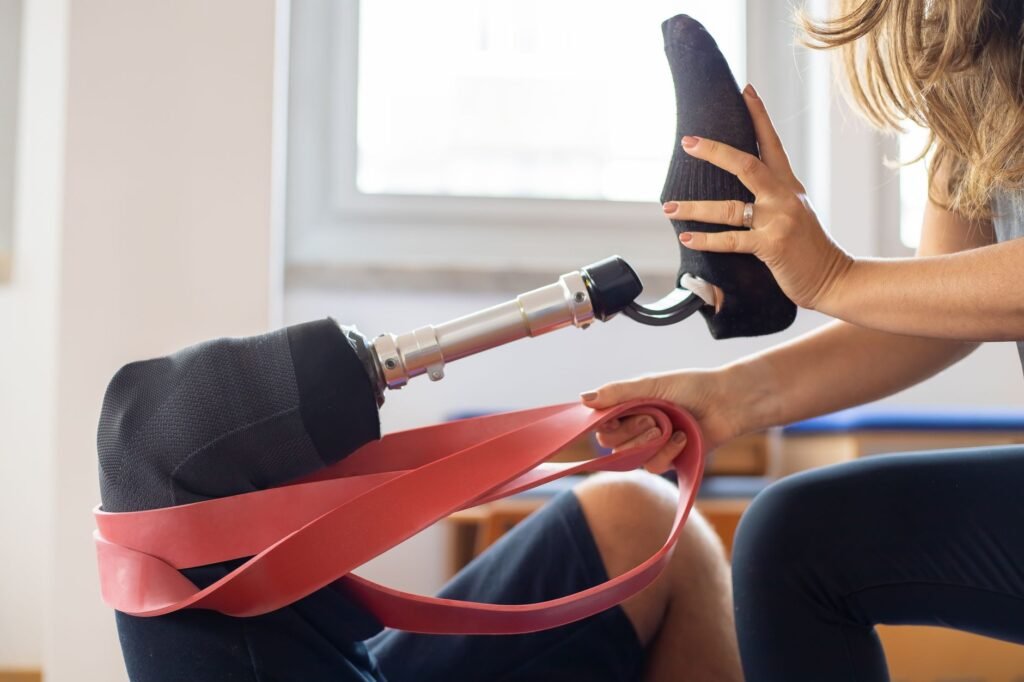
Therapists begin with pain‑free active range of motion exercises to restore joint glide without aggravating the bursa. Soft‑tissue mobilization and gentle myofascial release reduce surrounding muscle tension that can compress the bursa. Progressive resistance training targets the muscles that stabilize the affected joint, typically using elastic bands before advancing to free weights. Education on body mechanics helps patients avoid positions that previously triggered symptoms.
Gradual Loading Protocols
After pain subsides, clinicians introduce a structured loading schedule that increases stress in small increments, often 10% per week. Functional tasks such as stair climbing, light jogging, or sport‑specific drills are re‑introduced only after the patient can complete the previous step without pain. Monitoring of swelling and tenderness guides adjustments; setbacks are addressed by temporarily reducing load and adding restorative modalities.
Monitoring and Adjustments
Regular follow‑up appointments allow clinicians to assess bursal size with ultrasound and evaluate functional progress. If symptoms recur, a brief course of corticosteroid injection may be considered before modifying the rehabilitation plan. Patient‑reported outcome measures, such as the Visual Analogue Scale and the Disabilities of the Arm, Shoulder and Hand questionnaire, provide objective data to track recovery. Successful return to full activity is declared when the individual demonstrates pain‑free performance of sport or work tasks for at least two consecutive weeks.
- Start with pain‑free range of motion
- Progress resistance gradually
- Use objective outcome measures for tracking
- Adjust plan promptly if symptoms return
Frequently Asked Questions
Can bursitis become chronic?
Yes. Repeated irritation without adequate rest can lead to chronic inflammation, making symptoms last months or longer. Ongoing physical therapy and activity adjustments are key to breaking the cycle.
Is bursitis contagious?
Non‑infectious bursitis is not contagious. Only septic bursitis, caused by bacteria entering the bursa, involves an infection that could theoretically spread through direct wound contact, but it is rare.
Can I exercise with bursitis?
Low‑impact activities that do not stress the inflamed joint are generally safe. Swimming, stationary cycling, or walking can maintain fitness while allowing the bursa to heal. Always follow a therapist’s guidance and avoid painful movements.
What is the difference between bursitis and arthritis?
Both cause joint pain, but bursitis involves inflammation of the bursa, whereas arthritis is inflammation of the joint itself, affecting cartilage and bone. Symptoms can overlap, so professional assessment is important for accurate diagnosis.
The Bottom Line
Bursitis is a common, treatable condition that results from inflammation of the small fluid sacs cushioning our joints. Recognizing the signs, resting the affected area, using ice or heat, and taking appropriate over‑the‑counter medication usually leads to recovery within weeks. When symptoms linger or worsen, professional evaluation ensures proper treatment and rules out infection or other joint disorders. By combining medical care with sensible home strategies, most people return to their normal activities without lasting discomfort.
For more detailed information, you can visit MedlinePlus.
Was this page helpful?
Thanks for your feedback! What could we improve?
Thank you! Your feedback helps us improve our articles.
True Impact Health articles are written by our editorial team and based on reliable sources such as public health agencies, clinical guidelines and peer-reviewed research. This article has not yet been medically reviewed. Read our editorial process.
This content is for general information and is not a substitute for professional medical advice, diagnosis or treatment.