













Bone density test
Learn what a Bone density test is, who needs it, how it works, and how to understand the results for stronger, healthier bones.
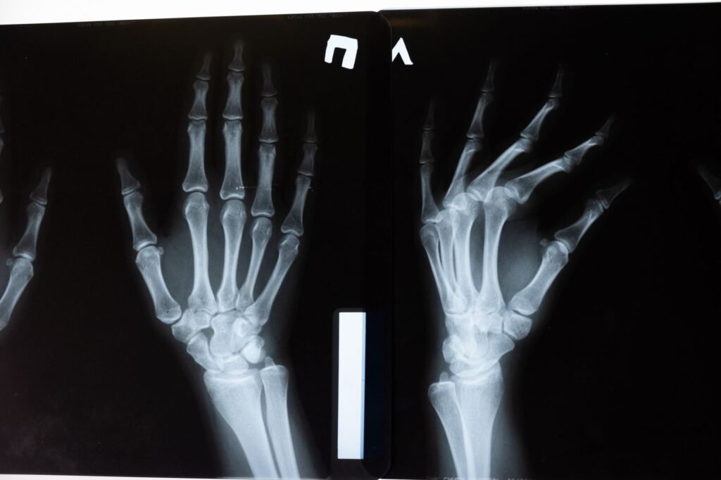
Table of Contents
Key Takeaways
- A bone density test measures bone strength and screens for osteoporosis.
- It is safe, quick, and recommended for people with specific risk factors.
- Results are reported as T‑scores and Z‑scores that guide treatment decisions.
- Preparation is simple: wear loose clothing and inform the clinic of any metal implants.
The Bone density test is a quick, painless way to see how strong your skeleton is and whether you are at risk for osteoporosis.
Many people hear the term and wonder if it is painful, expensive, or only for older women. In reality, the test is safe, widely available, and recommended for anyone with risk factors for bone loss.
Understanding what the test measures, who should have it, and how to interpret the results can empower you to take proactive steps for healthier bones.
What is a Bone Density Test?
Definition
A bone density test, also called a bone mineral density (BMD) test, uses low‑dose X‑ray technology to measure the amount of mineral (mostly calcium) in a specific area of bone. The most common method is dual‑energy X‑ray absorptiometry, known as DEXA or DXA.
How It Works
The DEXA machine sends two X‑ray beams at different energy levels through the bone. By comparing how much each beam is absorbed, the machine calculates bone density in grams per square centimeter.
The test focuses on the lumbar spine, hip, or sometimes the forearm because these sites are most predictive of fracture risk.
Why You Might Need One
Bone loss often occurs without obvious symptoms until a fracture happens. Detecting low bone density early lets you and your health professional intervene before a break occurs.
Guidelines from the National Osteoporosis Foundation and the Centers for Disease Control and Prevention recommend screening for:
- Women age 65 and older.
- Men age 70 and older.
- Post‑menopausal women under 65 with risk factors such as a family history of osteoporosis, low body weight, or prolonged steroid use.
- Adults of any age who have had a fracture after a minor fall.
For a clear overview of risk factors, you can visit MedlinePlus, a trusted health information site.
Even if you feel fine, a bone density test can reveal hidden weakness and guide lifestyle or medication choices that keep your skeleton strong.
How the Test Is Performed
The procedure takes about 10 to 20 minutes. You will lie on a padded table while the scanner moves over the area being examined. No contrast dye is needed.
Because the X‑ray dose is extremely low, much less than a standard chest X‑ray, there is no significant radiation risk for most people. However, pregnant women should discuss alternatives with their doctor.
Technicians may ask you to remove metal objects such as jewelry, belts, or hearing aids that could interfere with the image.
Understanding the Results
The DEXA report provides two key numbers: the T‑score and the Z‑score. Both compare your bone density to reference groups.
| Score | Interpretation |
|---|---|
| T‑score ≥ ‑1.0 | Normal bone density |
| T‑score between ‑1.0 and ‑2.5 | Low bone mass (osteopenia) |
| T‑score ≤ ‑2.5 | Osteoporosis |
The Z‑score compares your bone density to people of the same age, sex, and size. A Z‑score below ‑2.0 may suggest that factors other than aging are affecting bone health.
Doctors use these numbers along with your medical history to decide whether lifestyle changes, supplements, or prescription medication are needed.
Preparing for Your Appointment
Preparation is straightforward, but a few steps can improve accuracy and comfort:
- Wear loose‑fitting clothing without metal fasteners. A hospital gown is often provided.
- Tell the technician about any recent barium studies or contrast agents, as they can affect the scan.
- Bring a list of current medications, especially steroids, thyroid hormones, or bone‑active drugs.
- Schedule the test at the same time of day as any previous scans if you are monitoring changes over time.
Hydration is not a major factor, but staying comfortably hydrated can help you stay relaxed during the short procedure.

Who Should Get Tested
While age and gender are primary drivers, many medical conditions increase the need for early screening.
Medical Conditions
Conditions that can lower bone density include:
- Rheumatoid arthritis
- Chronic kidney disease
- Hyperthyroidism or hyperparathyroidism
- Eating disorders such as anorexia nervosa
Medications
Long‑term use of glucocorticoids (e.g., prednisone), certain anticonvulsants, and some cancer therapies can accelerate bone loss. Discuss with your doctor whether a test is warranted.
Lifestyle Factors
Low calcium intake, sedentary habits, smoking, and excessive alcohol consumption all contribute to weaker bones. If you have several of these risk factors, a bone density test may be advised even before the age thresholds.
Lifestyle Factors That Influence Bone Density
Nutrition Beyond Calcium
While calcium is often highlighted, a broader dietary pattern supports bone health. Adequate protein intake provides the amino acids needed for collagen formation, which forms the organic matrix of bone. Vitamin D from fortified foods or safe sun exposure enhances calcium absorption, and magnesium, potassium, and vitamin K2 contribute to mineralization and bone remodeling. Emphasizing a balanced diet reduces the need for supplementation alone.
Incorporating leafy greens, nuts, legumes, and fatty fish supplies these nutrients naturally. For individuals with restricted diets, a registered dietitian can tailor plans that meet bone‑supportive needs without excess calories. Consistency over months yields measurable improvements in bone turnover markers.
Physical Activity and Mechanical Loading
Weight‑bearing and resistance exercises generate mechanical strain that stimulates osteoblast activity. Walking, jogging, and stair climbing apply repetitive loading, while strength training with free weights or resistance bands targets specific skeletal sites such as the hip and spine. Research shows that regular activity can slow age‑related bone loss and, in some cases, increase bone mineral density.
Guidelines recommend at least 150 minutes of moderate aerobic activity combined with two sessions of muscle‑strengthening exercises each week. Progression should be gradual to avoid injury, and balance training can reduce fall risk, indirectly protecting bone integrity. A structured program under professional supervision maximizes benefit.
Smoking, Alcohol, and Other Modifiable Risks
Both tobacco use and excessive alcohol consumption negatively affect bone remodeling. Nicotine impairs osteoblast function and reduces calcium absorption, while chronic intake of more than two alcoholic drinks per day interferes with vitamin D metabolism. These behaviors also increase fall risk, compounding fracture potential.
Quitting smoking and limiting alcohol to moderate levels are practical steps that improve bone health outcomes. Support resources such as counseling, nicotine replacement, and community groups have demonstrated success. Integrating these changes with nutrition and exercise creates a comprehensive preventive strategy.
- Eat a diet rich in protein, leafy greens, nuts, and fatty fish.
- Engage in weight‑bearing activities most days of the week.
- Perform resistance training at least twice weekly.
- Avoid smoking and limit alcohol to no more than two drinks per day.

Medications and Their Impact on Bone Health
Drugs That Reduce Bone Density
Corticosteroids, often prescribed for inflammatory conditions, can accelerate bone loss by decreasing calcium absorption and increasing resorption. Long‑term use, especially at doses equivalent to prednisone 5 mg per day or higher, warrants proactive monitoring. Other agents such as certain anticonvulsants, proton pump inhibitors, and aromatase inhibitors also carry a risk of reduced bone mineral density.
Clinicians typically assess fracture risk before initiating these medications and may co‑prescribe bone‑protective agents when appropriate. Dose minimization, intermittent dosing schedules, and periodic drug holidays are strategies to mitigate adverse skeletal effects. Patients should be educated about the potential impact on bone health.
Bone‑Protective Pharmacotherapy
Bisphosphonates, including alendronate and risedronate, inhibit osteoclast‑mediated resorption and are first‑line treatments for osteoporosis. They have demonstrated reductions in vertebral and hip fracture rates in multiple randomized trials. Administration guidelines emphasize taking the medication with plain water, remaining upright for at least 30 minutes, and avoiding food for a short period to improve absorption.
Alternative agents such as denosumab, a monoclonal antibody targeting RANKL, provide a reversible option for patients who cannot tolerate oral bisphosphonates. Selective estrogen receptor modulators and hormone replacement therapy may be considered for postmenopausal women with specific risk profiles, but benefits must be weighed against cardiovascular and cancer risks. Regular follow‑up with bone density testing guides therapy duration.
Supplemental Interventions and Monitoring
Calcium and vitamin D supplementation remain foundational when dietary intake is insufficient, especially for patients on bone‑affecting medications. Typical recommendations include 1,000-1,200 mg of elemental calcium and 800-1,000 IU of vitamin D daily, adjusted for age, renal function, and baseline levels. Monitoring serum 25‑hydroxyvitamin D ensures adequacy and helps avoid toxicity.
When initiating pharmacologic therapy, baseline and follow‑up bone density scans are essential to assess response. Some clinicians also track biochemical markers such as serum C‑telopeptide or procollagen type 1 N‑terminal propeptide to gauge turnover. These measures complement imaging and inform adjustments in treatment plans.
- Identify medications that increase fracture risk.
- Consider bisphosphonates or denosumab for high‑risk patients.
- Supplement calcium (1,000‑1,200 mg) and vitamin D (800‑1,000 IU) as needed.
- Schedule baseline and periodic bone density assessments.
\n
Interpreting Bone Density Trends Over Time
Serial Scanning and Expected Changes
Repeated bone density measurements are typically performed every one to two years, depending on initial risk assessment and treatment status. Small changes may fall within the scanner’s precision error, so clinicians rely on the least significant change (LSC) to determine true progression or improvement. An increase exceeding the LSC suggests a positive response to therapy, whereas a decrease indicates potential treatment failure or emerging risk.
Age‑related decline varies by skeletal site; the lumbar spine often shows slower loss than the hip in postmenopausal women, while men may experience more uniform changes. Understanding these patterns helps avoid over‑interpretation of minor fluctuations. Consistency in positioning and equipment calibration further enhances reliability.
Integrating Clinical Context with Scan Results
Bone density numbers alone do not dictate management; they must be combined with clinical risk factors such as prior fractures, family history, glucocorticoid exposure, and fall propensity. Tools like FRAX incorporate both densitometric data and clinical variables to estimate 10‑year fracture probability. When the calculated risk surpasses treatment thresholds, pharmacologic intervention is generally recommended.
Patients with borderline scores may benefit from lifestyle optimization and close monitoring before initiating medication. Shared decision‑making, incorporating patient values and preferences, ensures adherence and satisfaction with the chosen plan. Documentation of risk factor changes over time supports dynamic adjustments to care.
Special Considerations for Different Populations
Individuals with chronic kidney disease, endocrine disorders, or secondary osteoporosis require tailored interpretation of bone density trends. For example, patients on dialysis may have altered bone turnover that is not fully captured by standard densitometry, prompting use of additional imaging or laboratory markers. Similarly, men with hypogonadism may need hormone replacement in conjunction with bone‑targeted therapy.
In pediatric and adolescent populations, growth and maturation affect bone density values, so age‑ and sex‑specific reference data are essential. Monitoring in these groups focuses on achieving peak bone mass rather than preventing fractures. Clinicians should be aware of these nuances to avoid misclassification.
- Schedule repeat scans every 1-2 years based on risk.
- Use least significant change to assess true differences.
- Combine densitometry with FRAX or similar tools.
- Adjust interpretation for comorbid conditions and age.
Frequently Asked Questions
Is the bone density test painful?
No. The test uses a low‑dose X‑ray beam that passes through the body. You may feel a brief pressure from the table, but there is no pain.
How often should I have the test?
For most adults, repeat testing every 2 to 5 years is sufficient. Your doctor may recommend more frequent scans if you have osteoporosis or are on medication that affects bone.
Can I do the test if I have a pacemaker?
Yes. The DEXA scan does not use magnetic fields, so it is safe for people with pacemakers or other implanted electronic devices.
Will my insurance cover the cost?
Many insurance plans, including Medicare Part B, cover the test when it is medically indicated. Check with your provider for specific coverage details.
The Bottom Line
A Bone density test is a simple, low‑risk way to assess bone health, identify osteoporosis early, and guide preventive actions. Whether you are approaching the recommended screening age or have risk factors, talking with your health professional about a DEXA scan can help keep your bones strong for years to come.
Was this page helpful?
Thanks for your feedback! What could we improve?
Thank you! Your feedback helps us improve our articles.
True Impact Health articles are written by our editorial team and based on reliable sources such as public health agencies, clinical guidelines and peer-reviewed research. This article has not yet been medically reviewed. Read our editorial process.
This content is for general information and is not a substitute for professional medical advice, diagnosis or treatment.