











Symptoms of perforated sinus after tooth extraction
Learn the key Symptoms of perforated sinus after tooth extraction, how to spot them early, and what steps to take for safe recovery.
By True Impact Health Editorial Team

Physician with experience in patient care and preventive medicine, focused on early detection, disease prevention and long-term wellness.
Reviewed this article on October 15, 2026.
View profile How we reviewUpdated on October 14, 2026 9 min read

Table of Contents
Key Takeaways
- A sinus perforation can occur during upper tooth extraction and often shows clear warning signs.
- Early symptoms include nasal discharge, foul taste, and facial pressure; seek dental care promptly.
- Imaging such as X‑ray or CT scan confirms the diagnosis and guides treatment.
- Proper aftercare and preventive techniques reduce the risk of future perforations.
When a routine dental procedure turns into an unexpected complication, the first thing you notice is often a change in how you feel. The phrase Symptoms of perforated sinus after tooth extraction may sound technical, but understanding it can help you act quickly and avoid further problems.
During an upper back tooth (premolar or molar) extraction, the thin bone that separates the tooth socket from the maxillary sinus can be breached. This creates a small opening, or perforation, that allows air, fluid, or even bacteria to move between the mouth and the sinus cavity. Recognizing the warning signs early can make a big difference in your comfort and recovery.
Understanding the Sinus and Tooth Connection
Anatomy of the Maxillary Sinus
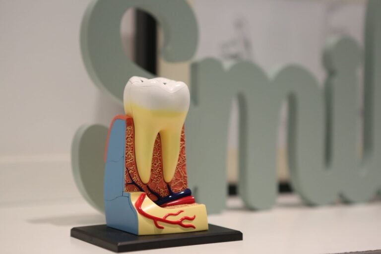
The maxillary sinus is a hollow, air‑filled space located behind the cheekbones and above the upper teeth. It is lined with a thin mucous membrane that produces mucus to keep the sinus moist and to trap particles.
In most adults, the floor of the sinus is only a few millimetres thick, especially near the roots of the second premolar and first molar. This close proximity is why dental extractions in that area carry a higher risk of creating a communication between the mouth and the sinus.
How Upper Back Teeth Relate to the Sinus
Upper premolars and molars often have roots that either touch or extend into the sinus floor. When a tooth is healthy, the bone provides a solid barrier. However, if the tooth is severely decayed, infected, or has been previously treated with a root canal, the surrounding bone may be weakened, making a perforation more likely during extraction.
Dental professionals assess this relationship with pre‑operative X‑rays or cone‑beam CT scans. Identifying a close root‑to‑sinus relationship ahead of time helps the dentist plan a careful approach and discuss potential risks with the patient.
What Is a Perforated Sinus?
Definition and Medical Context
A perforated sinus, also called an oro‑sinus communication, is a hole that forms between the oral cavity and the maxillary sinus. It is not a disease in itself but a pathway that can allow oral bacteria to enter the sinus, potentially leading to infection (sinusitis) or chronic drainage.
Most perforations are small, often just a few millimetres, and may close on their own with proper care. Larger openings may require surgical closure using a graft or a flap of tissue.
How Extraction Can Cause a Perforation
During extraction, the dentist uses elevators and forceps to loosen and remove the tooth. If excessive force is applied, or if the bone is already thin, the instrument can push through the sinus floor.
Complicating factors include:
- Previous infections that have eroded bone.
- Root morphology that is unusually long or curved.
- Inadequate imaging before the procedure.
Understanding these factors helps patients ask the right questions and understand why a perforation might happen.
Recognizing Early Warning Signs
Most people notice a change within the first few days after extraction. Early symptoms are usually mild but should never be ignored.
- Nasal discharge – clear or slightly cloudy fluid that drips from the nose, especially when leaning forward.
- Unusual taste – a salty or metallic taste in the mouth, often described as “mouth‑to‑nose” flavor.
- Facial pressure – a feeling of fullness or pressure in the cheek on the side of the extraction.
- Mild pain – throbbing pain that may worsen when you blow your nose or sneeze.
- Swelling – localized swelling around the extraction site that does not improve after 48 hours.
If any of these signs appear, contacting your dentist promptly can prevent the condition from worsening.
When Symptoms Escalate – Serious Indicators
While many perforations heal with conservative care, some develop complications that need urgent attention.
- Pus‑filled discharge – yellow or green mucus, sometimes with a foul odor, indicating infection.
- Fever – temperature above 38 °C (100.4 °F) suggests the body is fighting an infection.
- Severe facial pain – sharp, persistent pain that radiates to the eye or forehead.
- Difficulty breathing through the nose – blockage that does not improve with decongestants.
- Persistent bad taste or foul breath – may indicate ongoing drainage from the sinus into the mouth.
These signs warrant an immediate dental or medical evaluation to rule out acute sinusitis or a larger communication that may need surgical repair.
Diagnosis – How Dentists Confirm a Perforation
Diagnosis begins with a thorough clinical exam. The dentist will look for visible drainage, test the site with a blunt instrument, and listen for air bubbles when the patient gently blows their nose.
Imaging is the definitive tool. Common options include:
- Periapical X‑ray – provides a two‑dimensional view of the tooth roots and sinus floor.
- Panoramic radiograph – shows a broader view of the jaws and can reveal larger defects.
- Cone‑beam CT scan – offers three‑dimensional detail, allowing the dentist to measure the size of the perforation accurately.
For more information on imaging techniques, you can visit the MedlinePlus health information site.

Treatment Options and What to Expect
Treatment depends on the size of the perforation, presence of infection, and the patient’s overall health.
Conservative Management
Small perforations often heal on their own. The dentist may recommend:
- Keeping the extraction site clean with gentle saline rinses.
- Avoiding forceful nose blowing for at least 48 hours.
- Using a prescribed antibiotic if there are signs of infection.
- Applying a cold pack to reduce swelling.
Follow‑up visits are scheduled to monitor healing.
Surgical Closure
Larger or persistent openings may need a surgical procedure called an oro‑sinus membrane repair. Techniques include:
- Placement of a collagen membrane graft.
- Use of a buccal (cheek‑side) flap to cover the defect.
- Endoscopic sinus surgery in collaboration with an ENT specialist.
These procedures are usually performed under local anesthesia and have a high success rate.
Aftercare and Prevention Tips
Whether you receive conservative care or surgery, diligent aftercare reduces the chance of recurrence.
- Do not use a straw for the first week; suction can pull air into the sinus.
- Avoid smoking and exposure to second‑hand smoke, as it impairs healing.
- Maintain good oral hygiene with a soft‑bristled toothbrush and gentle rinses.
- Stay upright for the first 24 hours to limit blood pooling.
- Follow any prescribed medication schedule exactly as directed.
Managing Pain and Discomfort
Over-the-counter analgesics
Non‑prescription pain relievers are usually the first line of defense after a sinus perforation. Ibuprofen and naproxen reduce both pain and inflammation by inhibiting prostaglandin production. Acetaminophen can be added for patients who cannot tolerate non‑steroidal anti‑inflammatory drugs (NSAIDs). It is important to follow dosing instructions and avoid exceeding the maximum daily limit.
- Ibuprofen 200‑400 mg every 6‑8 hours
- Naproxen 250‑500 mg every 12 hours
- Acetaminophen 500‑1000 mg every 6 hours
Cold and heat therapy
Applying a cold pack to the cheek for 15 minutes can constrict blood vessels and lessen swelling during the first 24‑48 hours. After the initial period, a warm compress may improve sinus drainage and relieve lingering pressure. Alternate between cold and heat only if the skin tolerates the temperature changes and avoid direct contact with ice.
- Cold pack: 15 minutes on, 15 minutes off, repeat up to 4 times daily
- Warm compress: 10‑15 minutes, 2‑3 times daily after 48 hours

Impact on Dental Restorations and Implants
Effect on crowns and bridges
A perforated sinus can compromise the support needed for fixed prostheses placed near the maxillary molars. The loss of bone height may require a temporary reduction in occlusal load to prevent further displacement. Dentists often recommend a provisional restoration that avoids direct pressure on the affected site while healing progresses.
- Use a temporary crown with reduced thickness
- Limit chewing on the side of the perforation for 2‑3 weeks
- Schedule a follow‑up radiograph to assess bone regeneration
Implant planning after sinus involvement
When an implant is planned in the posterior maxilla, a healed sinus membrane is essential for predictable osseointegration. If a perforation occurs, a sinus lift may be postponed until the membrane re‑epithelializes, typically 4‑6 weeks. In some cases, a graft material is placed simultaneously with the membrane repair to preserve ridge height.
- Delay definitive implant placement until imaging confirms membrane integrity
- Consider using short or tilted implants if bone height remains limited
- Coordinate with an oral surgeon experienced in sinus augmentation
When to Seek Emergency Care
Signs of infection spreading
Although mild swelling is common, rapid expansion of facial swelling, fever above 38 °C, or a foul‑smelling discharge from the extraction site signal a possible infection that has entered the sinus cavity. Prompt medical evaluation is needed to prevent cellulitis or orbital involvement. Antibiotic therapy may be escalated based on culture results.
- Fever or chills
- Increasing facial or periorbital swelling
- Purulent nasal discharge or foul taste
Severe bleeding or sudden vision changes
Heavy bleeding that does not stop with pressure, or any sudden change in vision such as double vision or loss of visual acuity, requires immediate attention. These symptoms can indicate vascular injury or extension of the perforation into the orbital floor. Call emergency services or go to the nearest emergency department without delay.
- Uncontrolled oral or nasal bleeding
- Blurred or double vision
- Sudden loss of sight in one eye
Frequently Asked Questions
Can a perforated sinus heal without surgery?
Yes, many small perforations close on their own within a few weeks if the area is kept clean and the patient avoids activities that increase sinus pressure.
How long does it take to recover from a surgical repair?
Most patients feel comfortable enough to resume normal activities within 5‑7 days, but full healing of the sinus lining can take up to 6‑8 weeks.
Is it safe to take over‑the‑counter pain relievers?
Acetaminophen is generally safe. Non‑steroidal anti‑inflammatory drugs (NSAIDs) like ibuprofen are also fine unless you have specific contraindications. Always check with your dentist or physician.
Will a perforated sinus affect future dental work?
Once healed, the sinus floor regains its strength. However, dentists may take extra imaging before any future extractions or implants in the same area to ensure the bone is sufficient.
The Bottom Line
Recognizing the Symptoms of perforated sinus after tooth extraction early can prevent infection and reduce the need for surgery. Simple signs such as nasal discharge, unusual taste, and facial pressure should prompt a call to your dentist. With prompt diagnosis, most perforations heal quickly, and careful aftercare helps you get back to feeling normal.
Was this page helpful?
Thanks for your feedback! What could we improve?
Thank you! Your feedback helps us improve our articles.
Medically reviewed by
Dr. Bernard ReynoldsPhysician with experience in patient care and preventive medicine, focused on early detection, disease prevention and long-term wellness.
True Impact Health articles are written by our editorial team and based on reliable sources such as public health agencies, clinical guidelines and peer-reviewed research. This article has been checked by a doctor on our review panel. Read our editorial process.
This content is for general information and is not a substitute for professional medical advice, diagnosis or treatment.