Introduction
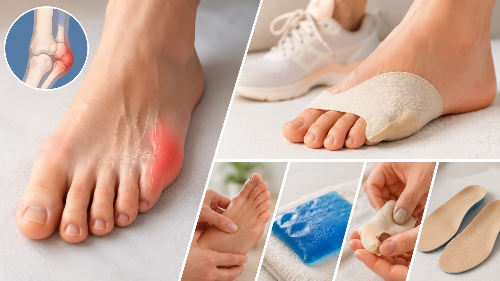
A tailor’s bunion, also known as a bunionette, is a common foot condition that develops on the outside of the foot at the base of the little toe. Although it is generally smaller than a traditional bunion that forms near the big toe, it can still cause significant pain, swelling, and difficulty wearing shoes comfortably. Many people initially ignore the condition, assuming it is simply a callus or minor irritation. However, without proper management, a tailor’s bunion can gradually worsen and interfere with daily activities, exercise, and overall quality of life.
Fortunately, most cases can be successfully managed through conservative treatment approaches such as footwear modifications, pain relief measures, targeted exercises, and lifestyle adjustments. In more advanced situations, surgical intervention may be necessary to correct the underlying structural problem. Understanding the causes, symptoms, treatment options, and prevention strategies can help you take control of the condition before it becomes debilitating. This comprehensive guide explains everything you need to know about treating a tailor’s bunion and maintaining healthy, pain-free feet.
Understanding Tailor’s Bunion
What Is a Tailor’s Bunion?
A tailor’s bunion is a bony enlargement that develops at the joint where the fifth metatarsal bone meets the little toe. The fifth metatarsal is the long bone located on the outer side of the foot. When this bone gradually shifts outward or becomes more prominent, a noticeable bump forms on the outside edge of the foot.
As the deformity progresses, pressure from footwear and daily walking can irritate the surrounding tissues. This often leads to inflammation, redness, tenderness, and increasing discomfort. In some cases, the little toe may also begin to angle inward toward the other toes, further contributing to pain and shoe-fitting difficulties.
Why Is It Called a Tailor’s Bunion?
The condition earned its name centuries ago when tailors commonly sat cross-legged for long periods while sewing garments. This sitting position placed constant pressure on the outer edges of their feet, particularly near the fifth metatarsal bone. Over time, many tailors developed these characteristic bumps, leading to the term “tailor’s bunion.”
Although modern lifestyles have changed significantly, the condition remains common due to footwear choices, inherited foot structures, and repetitive stress on the feet.
Related Posts:


Common Causes and Risk Factors
Several factors can contribute to the development of a tailor’s bunion. In many cases, multiple factors work together to increase the likelihood of the condition.
Foot Structure and Biomechanics
Natural foot anatomy plays a major role in bunionette formation. Individuals with unusually high arches, flat feet, or abnormal foot mechanics may place excessive pressure on the outer edge of the foot while walking. A naturally enlarged or outwardly angled fifth metatarsal bone can also increase the risk.
When weight is distributed unevenly across the foot, repetitive stress gradually causes the joint to protrude outward, eventually forming a bunionette.
Improper Footwear
One of the most common contributing factors is wearing shoes that compress the forefoot. Narrow shoes, pointed-toe footwear, and high heels force the toes into unnatural positions and increase pressure on the little toe joint.
Over time, this constant pressure irritates the tissues around the fifth metatarsal and accelerates deformity development. Even individuals with a genetic predisposition may avoid significant symptoms if they consistently wear properly fitting footwear.
Genetics and Family History
Many people inherit foot structures that make them more susceptible to bunions and tailor’s bunions. If close family members have experienced bunionettes, there is a greater chance of developing the condition yourself.
Inherited characteristics such as foot shape, bone alignment, ligament laxity, and gait patterns can all contribute to risk.
Repetitive Activities and Injuries
Certain sports and activities place repeated stress on the outer edge of the foot. Running, dancing, hiking, and occupations that require prolonged standing may increase pressure on the fifth metatarsal joint.
Previous foot injuries may also alter walking mechanics and contribute to bunionette formation.
Arthritis and Inflammatory Conditions
Inflammatory disorders such as rheumatoid arthritis can weaken joints and contribute to bone deformities. Chronic inflammation may accelerate the progression of a tailor’s bunion and increase pain levels.
Recognizing the Symptoms
Early identification of symptoms allows for earlier treatment and may help prevent progression.
The most noticeable symptom is a visible bump on the outside of the foot near the base of the little toe. Initially, this bump may be small and painless. As the condition worsens, it often becomes tender and inflamed.
Many individuals experience redness and swelling around the affected joint, particularly after long periods of walking or standing. Friction from shoes can lead to the formation of calluses or corns over the prominence.
Pain is another common complaint. Some people experience discomfort only when wearing certain shoes, while others report persistent aching that interferes with daily activities. Walking, exercising, and standing for extended periods may become increasingly difficult.
In advanced cases, finding comfortable footwear becomes challenging because the enlarged joint rubs against the inside of shoes.
Non-Surgical Treatment Options
Fortunately, most tailor’s bunions can be treated effectively without surgery. Conservative treatment focuses on reducing pressure, relieving pain, and preventing further progression.
Footwear Modifications
Changing footwear is often the most important first step in treatment.
Shoes with wide toe boxes allow the toes to spread naturally and reduce pressure on the affected joint. Soft leather or mesh materials can also minimize irritation.
Low-heeled shoes with adequate cushioning and arch support help distribute weight more evenly across the foot. Avoiding pointed shoes and high heels can significantly reduce symptoms and slow progression.
Many patients experience noticeable improvement simply by switching to properly fitted footwear.
Orthotics and Padding
Custom orthotics can help correct abnormal foot mechanics and redistribute pressure away from the fifth metatarsal. These devices are especially beneficial for individuals with flat feet, high arches, or gait abnormalities.
Protective bunion pads, silicone sleeves, and cushioning inserts can further reduce friction and discomfort when wearing shoes.
Ice Therapy
Applying ice packs to the affected area can help reduce inflammation and pain. Ice should be applied for approximately 10 to 15 minutes at a time, several times per day, particularly after physical activity.
Wrapping the ice pack in a towel helps prevent skin irritation and frostbite.
Pain Relief Medications
Over-the-counter nonsteroidal anti-inflammatory drugs (NSAIDs), such as ibuprofen or naproxen, may provide temporary relief from pain and swelling.
Topical anti-inflammatory gels and pain-relieving creams can also help manage symptoms without the systemic side effects associated with oral medications.
Individuals with underlying medical conditions should consult their healthcare provider before using NSAIDs regularly.
Exercises for Tailor’s Bunion
Targeted exercises can improve foot flexibility, strengthen supportive muscles, and reduce stress on the affected joint.
Toe Stretching Exercises
Gently pulling the toes backward helps improve joint mobility and relieve stiffness. Holding the stretch for 20 to 30 seconds several times daily may improve flexibility and comfort.
Towel Curls
Placing a towel on the floor and using the toes to pull it toward the body strengthens the intrinsic muscles of the foot. This exercise helps support proper foot mechanics and improves stability.
Marble Pickups
Picking up small objects such as marbles with the toes enhances coordination and strengthens foot muscles. This simple exercise can be performed while sitting and requires only a few minutes each day.
Calf Stretching
Tight calf muscles can alter walking mechanics and increase pressure on the forefoot. Regular calf stretches help improve overall lower-extremity flexibility and reduce strain on the foot.
Physical Therapy and Professional Treatment
When symptoms persist despite home treatment, physical therapy may provide additional benefits.
A physical therapist or podiatrist can evaluate walking patterns, identify biomechanical abnormalities, and develop a personalized treatment plan. Manual therapy techniques, stretching programs, strengthening exercises, and gait retraining may all contribute to symptom relief.
Some practitioners also use ultrasound therapy, laser therapy, or other modalities to reduce inflammation and promote healing.
When Surgery May Be Necessary
Surgery is generally reserved for patients who continue to experience significant pain despite comprehensive conservative treatment.
Distal Fifth Metatarsal Osteotomy
This commonly performed procedure involves cutting and realigning the fifth metatarsal bone closer to the affected joint. The bone is then stabilized with screws or other fixation devices.
Proximal Osteotomy
For more severe deformities, realignment may be performed closer to the middle of the foot. This approach allows for greater correction of the underlying structural problem.
Exostectomy
In selected cases, the surgeon may simply remove the bony prominence. However, because this procedure does not address the underlying alignment issue, it is often combined with additional corrective techniques.
Soft Tissue Procedures
Tight ligaments, tendons, and joint capsules may be released or repositioned to improve alignment and reduce pressure on the affected area.
Recovery After Surgery
Recovery varies depending on the specific procedure performed.
Most patients wear a protective surgical shoe or walking boot for several weeks following surgery. Swelling and discomfort are expected during the early healing phase.
Gradual weight-bearing is typically introduced according to the surgeon’s recommendations. Physical therapy may be prescribed to restore mobility, strength, and normal walking patterns.
Most individuals return to normal daily activities within two to three months, although complete recovery may take longer in more complex cases.
Preventing Tailor’s Bunions
While not all cases can be prevented, several strategies can reduce risk and minimize recurrence.
Choosing shoes with adequate width, cushioning, and support remains one of the most effective preventive measures. Footwear should allow enough room for natural toe movement without excessive pressure.
Maintaining a healthy body weight reduces stress on the feet and lowers the risk of structural changes over time.
Regular foot exercises help maintain flexibility, strength, and proper alignment. Individuals with known foot abnormalities may benefit from custom orthotics to optimize weight distribution.
Routine foot inspections can also help identify early signs of irritation or deformity before symptoms become severe.
Conclusion
A tailor’s bunion may begin as a minor annoyance, but without proper attention it can gradually become a significant source of pain and mobility limitations. Fortunately, most cases respond well to conservative treatments such as wider footwear, orthotics, pain management strategies, and targeted exercises.
Early intervention is essential. Addressing symptoms promptly can slow progression, reduce discomfort, and help avoid the need for surgery. For individuals with persistent pain or advanced deformities, surgical correction can provide long-term relief and improved function.
By understanding the causes of tailor’s bunions and adopting proactive foot-care habits, you can protect your feet, maintain mobility, and continue enjoying an active, comfortable lifestyle for years to come.
Disclaimer
This article is for educational purposes only and does not replace professional medical advice, diagnosis, or treatment. Seizures, severe headache, confusion, visual changes, or blood pressure readings in the hypertensive crisis range require urgent medical evaluation.