
Introduction
Intracranial hypertension (IH) is a neurological condition characterized by elevated pressure within the skull. Because the skull is a rigid structure that cannot expand, any increase in pressure can affect the brain, optic nerves, blood vessels, and cerebrospinal fluid circulation. If left untreated, intracranial hypertension can lead to persistent headaches, visual disturbances, cognitive difficulties, and, in severe cases, permanent vision loss or neurological damage.
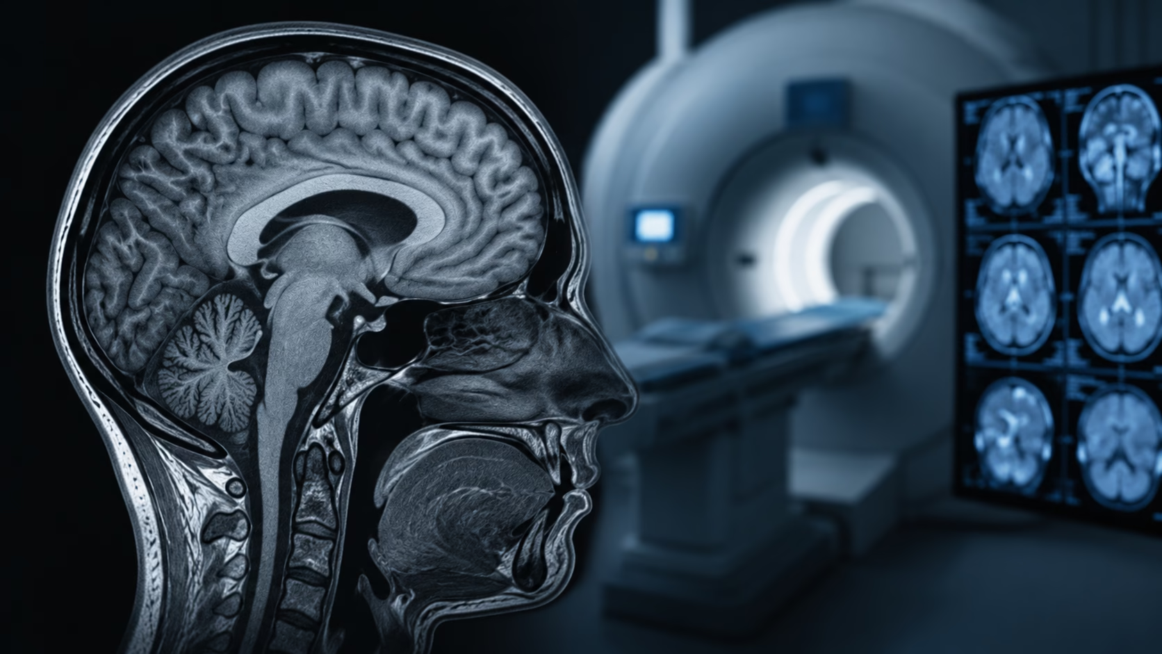
Diagnosing intracranial hypertension requires a combination of clinical evaluation, imaging studies, and sometimes lumbar puncture measurements. Among the available imaging techniques, Magnetic Resonance Imaging (MRI) has become one of the most valuable diagnostic tools. MRI provides detailed visualization of brain structures, cerebrospinal fluid spaces, and blood vessels without exposing patients to ionizing radiation. It helps clinicians identify characteristic signs of elevated intracranial pressure while also ruling out potentially life-threatening causes such as tumors, hemorrhages, or venous thrombosis.
Understanding how MRI contributes to the diagnosis and management of intracranial hypertension can help patients and healthcare providers appreciate its critical role in preventing complications and guiding treatment decisions.
Understanding Intracranial Hypertension
Intracranial pressure refers to the pressure exerted by brain tissue, blood, and cerebrospinal fluid within the skull. Under normal circumstances, intracranial pressure remains within a tightly regulated range, generally between 5 and 15 mmHg in adults. When this pressure rises beyond normal limits, intracranial hypertension develops.
Intracranial hypertension can occur for many reasons. In some patients, the condition develops without an identifiable cause and is referred to as idiopathic intracranial hypertension (IIH). This form is most commonly seen in women of childbearing age and is often associated with obesity. In other cases, elevated intracranial pressure develops secondary to underlying conditions such as brain tumors, traumatic brain injuries, hydrocephalus, infections, intracranial hemorrhage, or venous sinus thrombosis.
The symptoms of intracranial hypertension can vary from person to person. Persistent headaches are among the most common complaints and are often worse in the morning or when lying down. Many patients also experience visual disturbances, including blurred vision, double vision, transient visual obscurations, or loss of peripheral vision. Additional symptoms may include nausea, vomiting, pulsatile tinnitus, dizziness, and difficulty concentrating.
Related Posts:


Because these symptoms overlap with many other neurological conditions, accurate diagnosis is essential. MRI serves as a crucial component of that diagnostic process.
Why Early Detection Is Important
Early recognition and diagnosis of intracranial hypertension are essential because prolonged elevation of intracranial pressure can cause irreversible damage. One of the greatest concerns is injury to the optic nerves. Increased pressure can compress the optic nerve and lead to papilledema, a swelling of the optic disc that may eventually result in permanent vision loss if not treated promptly.
In addition to visual complications, chronic intracranial hypertension can interfere with normal brain function, leading to persistent headaches, cognitive impairment, and reduced quality of life. Identifying the condition early allows healthcare providers to implement treatments that reduce pressure, preserve vision, and prevent long-term neurological complications.
MRI plays a central role in this early detection process by revealing structural and vascular abnormalities that may otherwise remain undetected.
The Role of MRI in Diagnosing Intracranial Hypertension
Magnetic Resonance Imaging is considered one of the most valuable imaging modalities for evaluating patients with suspected intracranial hypertension. Unlike computed tomography (CT), MRI provides exceptional soft tissue detail and allows clinicians to visualize subtle anatomical changes associated with elevated intracranial pressure.
One of the primary advantages of MRI is its ability to identify secondary causes of intracranial hypertension. Brain tumors, cysts, hydrocephalus, inflammatory conditions, hemorrhages, and vascular abnormalities can all contribute to increased intracranial pressure. MRI helps clinicians detect or exclude these conditions before confirming a diagnosis of idiopathic intracranial hypertension.
Another major benefit is that MRI does not use ionizing radiation. This makes it particularly suitable for younger patients and those who require repeated imaging over time to monitor disease progression or treatment response.
MRI also provides detailed information about the brain, optic nerves, pituitary gland, cerebrospinal fluid spaces, and venous drainage pathways, all of which can be affected by elevated intracranial pressure.
Important MRI Sequences Used in Intracranial Hypertension
A comprehensive MRI examination for intracranial hypertension typically includes several specialized imaging sequences, each serving a unique diagnostic purpose.
T1-Weighted Imaging
T1-weighted sequences provide excellent anatomical detail and allow radiologists to evaluate brain structures with high precision. These images are particularly useful for assessing the pituitary gland and identifying an empty sella, a common finding in intracranial hypertension.
An empty sella occurs when elevated cerebrospinal fluid pressure compresses the pituitary gland, causing it to appear flattened against the floor of the sella turcica. Although not specific to intracranial hypertension, this finding frequently supports the diagnosis when combined with other imaging features.
T2-Weighted and FLAIR Imaging
T2-weighted and Fluid-Attenuated Inversion Recovery (FLAIR) sequences highlight fluid-containing structures and are useful for identifying subtle brain abnormalities. These images help evaluate ventricular size, white matter changes, and areas of edema.
In patients with intracranial hypertension, T2-weighted imaging often demonstrates enlargement of the optic nerve sheath and other cerebrospinal fluid-related changes that reflect increased pressure within the cranial compartment.
MR Venography (MRV)
Magnetic Resonance Venography is an essential component of many intracranial hypertension evaluations. MRV specifically examines the venous drainage pathways within the brain.
One of the most common findings in idiopathic intracranial hypertension is narrowing of the transverse venous sinuses. MRV can identify these areas of stenosis and help differentiate idiopathic intracranial hypertension from cerebral venous sinus thrombosis, a potentially serious condition that can mimic many of the same symptoms.
Because treatment strategies differ significantly between these disorders, MRV plays an important role in accurate diagnosis.
Orbital MRI Sequences
Specialized orbital imaging focuses on the eyes and optic nerves. High-resolution T2-weighted and fat-suppressed sequences provide detailed visualization of the optic nerve sheath and surrounding structures.
These images can reveal enlargement of the optic nerve sheath, protrusion of the optic nerve head, and flattening of the posterior globe, all of which are classic signs of elevated intracranial pressure.
Orbital imaging is particularly valuable in patients who present with visual symptoms or documented papilledema.
Phase-Contrast CSF Flow Studies
Phase-contrast MRI allows clinicians to evaluate cerebrospinal fluid movement and flow dynamics. Although not routinely performed in every case, these studies can provide additional information regarding cerebrospinal fluid circulation and pressure-related abnormalities.
CSF flow studies may be particularly helpful when differentiating intracranial hypertension from other neurological conditions that cause headaches or visual disturbances.
Characteristic MRI Findings in Intracranial Hypertension
Several imaging features are commonly associated with elevated intracranial pressure. While no single finding is diagnostic on its own, the presence of multiple characteristic abnormalities strongly supports the diagnosis.
Empty Sella
An empty or partially empty sella is one of the most frequently observed MRI findings in idiopathic intracranial hypertension. Increased cerebrospinal fluid pressure compresses the pituitary gland, causing it to appear flattened within the sella turcica.
Optic Nerve Sheath Enlargement
The optic nerve is surrounded by cerebrospinal fluid. Elevated intracranial pressure can cause distension of the optic nerve sheath, which appears enlarged on MRI.
This finding correlates with increased pressure transmission to the optic nerve and may contribute to visual symptoms.
Posterior Globe Flattening
Elevated pressure behind the eye can flatten the posterior aspect of the globe. This feature is highly suggestive of intracranial hypertension and is often identified during orbital MRI evaluation.
Optic Nerve Head Protrusion
Pressure-related swelling of the optic disc may cause protrusion of the optic nerve head into the globe. This imaging finding corresponds to papilledema observed during ophthalmologic examination.
Transverse Sinus Stenosis
Narrowing of the transverse venous sinuses is frequently identified in patients with idiopathic intracranial hypertension. Although the exact relationship remains complex, venous sinus stenosis may contribute to impaired cerebrospinal fluid drainage and elevated intracranial pressure.
Interpreting MRI Results
MRI findings should always be interpreted in conjunction with clinical symptoms, ophthalmologic examinations, and lumbar puncture results when available.
Radiologists typically evaluate the presence and severity of imaging abnormalities and compare current studies with previous examinations whenever possible. Standardized reporting helps ensure clear communication among neurologists, ophthalmologists, neurosurgeons, and primary care providers.
A diagnosis of intracranial hypertension should never rely solely on imaging findings. Instead, MRI serves as one important piece of a comprehensive diagnostic evaluation.
Patient Preparation and Safety Considerations
MRI is generally a safe and noninvasive procedure, but certain precautions are necessary before imaging begins.
Patients are screened for metallic implants, pacemakers, aneurysm clips, cochlear implants, and other devices that may be incompatible with MRI. Individuals with severe claustrophobia should inform their healthcare provider beforehand, as mild sedation or open MRI systems may improve comfort during the examination.
Most MRI studies for intracranial hypertension do not require special preparation. However, if contrast material is needed, patients may undergo kidney function testing beforehand to ensure safe administration.
How MRI Guides Treatment Decisions
MRI findings play a critical role in determining the most appropriate treatment strategy. By ruling out secondary causes and identifying characteristic features of intracranial hypertension, MRI helps clinicians develop personalized management plans.
For patients with idiopathic intracranial hypertension, treatment may include weight loss, medications such as acetazolamide, or therapeutic lumbar punctures to reduce pressure. In more severe cases, surgical interventions such as cerebrospinal fluid shunting or venous sinus stenting may be considered.
MRI also provides a valuable baseline for future comparison. Follow-up imaging can help assess treatment response, monitor disease progression, and identify complications that may require additional intervention.
Follow-Up Imaging
Patients diagnosed with intracranial hypertension often undergo repeat MRI or MR venography studies during follow-up care. Imaging intervals vary depending on symptom severity, visual status, and treatment response.
Many specialists recommend repeat imaging approximately six to twelve months after initiating treatment. Earlier imaging may be necessary if symptoms worsen, vision deteriorates, or new neurological deficits develop.
Regular monitoring helps ensure that treatment remains effective and that complications are detected promptly.
Conclusion
Magnetic Resonance Imaging has become an indispensable tool in the evaluation of intracranial hypertension. By providing detailed visualization of brain structures, optic nerves, cerebrospinal fluid spaces, and venous drainage pathways, MRI helps clinicians diagnose elevated intracranial pressure, identify underlying causes, and guide treatment decisions.
Characteristic findings such as empty sella, optic nerve sheath enlargement, posterior globe flattening, and transverse sinus stenosis can provide valuable diagnostic clues when interpreted alongside clinical and ophthalmologic findings. Furthermore, MRI allows physicians to monitor treatment effectiveness and track disease progression without exposing patients to radiation.
For patients experiencing persistent headaches, visual disturbances, or other symptoms suggestive of intracranial hypertension, timely MRI evaluation can make a significant difference. Early diagnosis and appropriate management not only improve symptoms but also help preserve vision and prevent long-term neurological complications. As imaging technology continues to advance, MRI will remain at the forefront of diagnosing and managing this important neurological condition.
Disclaimer
This article is for educational purposes only and does not replace professional medical advice, diagnosis, or treatment. Seizures, severe headache, confusion, visual changes, or blood pressure readings in the hypertensive crisis range require urgent medical evaluation.