






Fractures
Learn what fractures are, how they happen, symptoms, diagnosis, treatment and recovery tips in this clear, friendly guide for everyday readers.
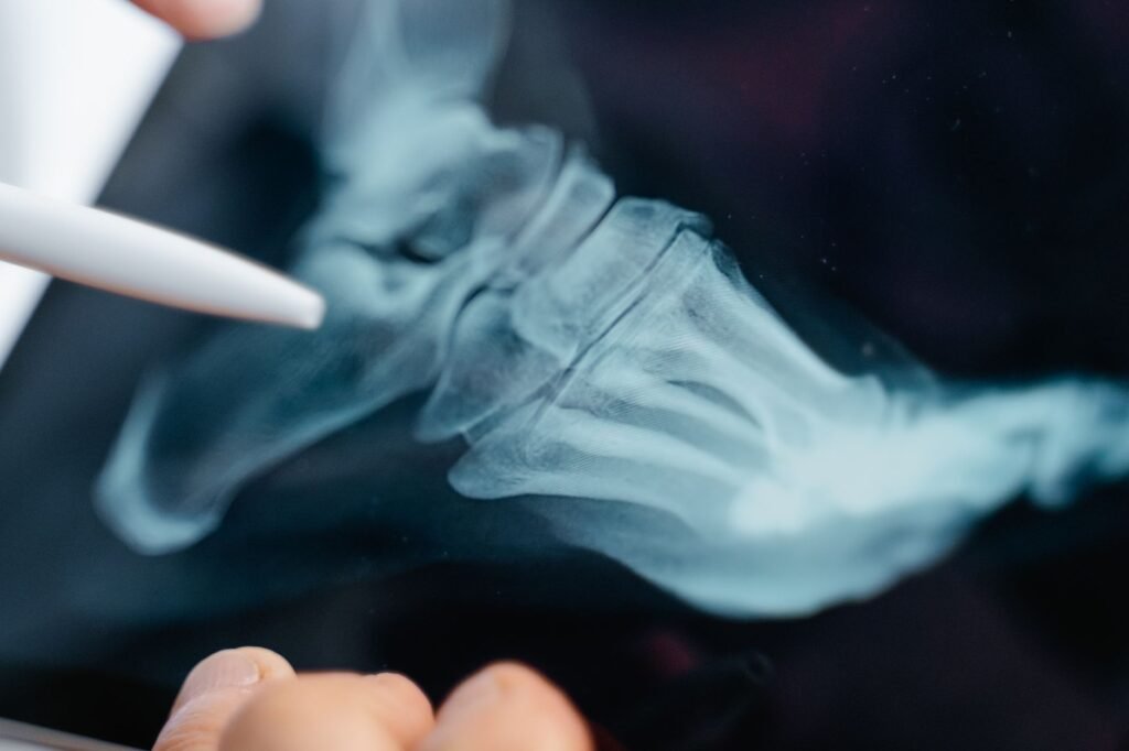
Table of Contents
Key Takeaways
- Fractures are breaks in bone that need prompt medical attention
- Common types include hairline, displaced, and compound fractures
- Treatment ranges from casting to surgery depending on severity
- Rehabilitation focuses on safe movement, nutrition and bone health
Fractures are a common injury that can happen to anyone, from a child falling off a bike to an older adult slipping on ice. Understanding what a fracture is, how to recognize it, and what to do next can make a big difference in healing and comfort.
This guide walks you through the basics of fractures, the different types, why they occur, how they are diagnosed, and what treatment options are available. It also offers practical advice for recovery, so you can return to daily life safely.
Remember, while this information is reliable and based on trusted sources such as the MedlinePlus website, it does not replace professional medical advice. If you suspect a fracture, seek care from a qualified health professional promptly.
What Is a Fracture?
A fracture is a break in the continuity of a bone. Bones are living tissue that can bend and flex, but when a force exceeds their strength, a crack or break occurs. The severity can range from a tiny hair‑line crack to a complete break that pierces the skin.
Fractures are classified by their shape, location, and whether the bone fragments stay aligned. The classification helps doctors decide the best treatment plan.
Most fractures heal with proper care, but complications such as infection, misalignment, or delayed healing can arise, especially if treatment is delayed.
Common Types of Fractures
Understanding the different ways a bone can break helps you recognize the injury and communicate clearly with health professionals.
Hairline (Stress) Fracture
This is a thin crack in the bone that often results from repetitive stress, such as running long distances or overuse in sports. Pain may be mild at first and worsen with activity.
Transverse Fracture
A straight‑across break caused by a direct blow or a sudden twist. The bone fragments usually stay in line, making casting an effective treatment.
Oblique Fracture
Angled break that occurs when a force hits the bone at an angle. These may shift more easily, sometimes requiring a splint or surgery.
Comminuted Fracture
The bone shatters into three or more pieces. This type often results from high‑impact injuries like car accidents and may need surgical fixation.
Compound (Open) Fracture
Here the broken bone pierces the skin, creating an open wound. This type carries a higher infection risk and always requires urgent medical care.
Greenstick Fracture
Common in children whose bones are more flexible. The bone bends and cracks on one side, similar to a green twig.
| Fracture Type | Typical Cause | Common Location |
|---|---|---|
| Hairline | Repetitive stress | Metatarsals, tibia |
| Transverse | Direct blow | Radius, femur |
| Oblique | Angled impact | Ulna, humerus |
| Comminuted | High‑impact trauma | Pelvis, tibia |
| Compound | Severe crush | Any bone, often tibia |
| Greenstick | Flexion injury | Forearm in children |
Causes and Risk Factors
Fractures can happen to anyone, but certain factors increase the likelihood of a break.
- Age – Bones become more brittle with age, especially after menopause.
- Osteoporosis – Reduced bone density makes fractures more likely from minor falls.
- High‑impact activities – Sports like football, skiing, or motorcycling.
- Falls – Common in older adults and children learning to walk.
- Weak muscles – Poor muscle support can put extra stress on bones.
Other contributors include certain medications (like long‑term steroids), nutritional deficiencies (especially calcium and vitamin D), and chronic conditions such as rheumatoid arthritis.
Reducing risk involves regular weight‑bearing exercise, a balanced diet rich in calcium, and safety measures like wearing helmets and using handrails.
Signs and Symptoms
Recognizing a fracture early can speed up treatment and improve outcomes. Typical signs include:
- Severe pain at the injury site, especially when moving the limb.
- Swelling, bruising, or tenderness.
- Deformity – the bone may look out of place or bent.
- Difficulty bearing weight or using the affected part.
- Visible bone protruding through the skin (in compound fractures).
Sometimes a fracture may be subtle, with only mild pain and swelling. If you suspect a break, it is safest to treat it as a fracture until a professional evaluates it.
Diagnosis
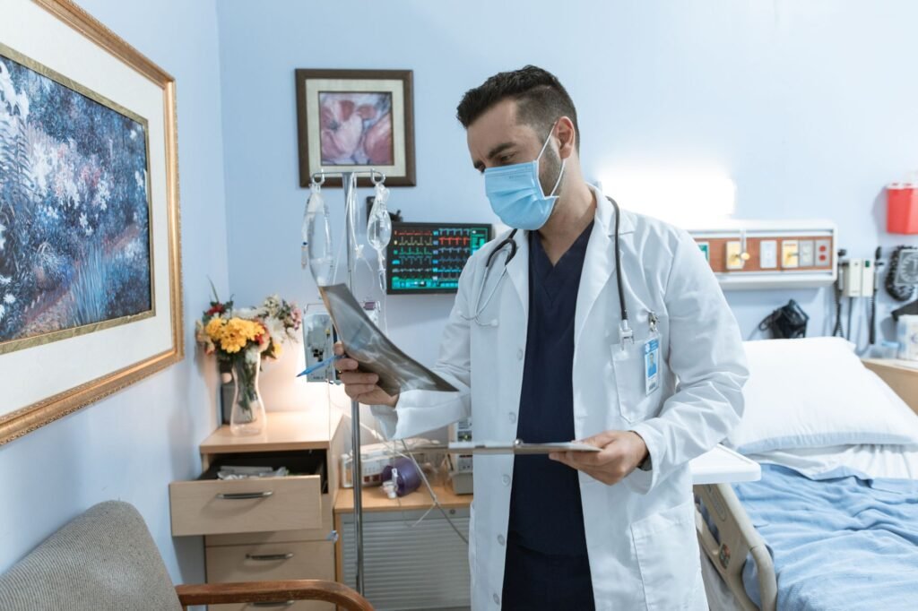
Medical professionals use a combination of physical examination and imaging to confirm a fracture.
Physical Examination
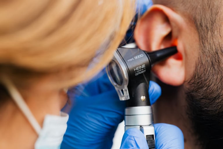
The doctor will gently palpate the area, assess range of motion, and look for signs of deformity or open wounds. They may also check for neurovascular status – ensuring nerves and blood flow are intact.
Imaging Tests
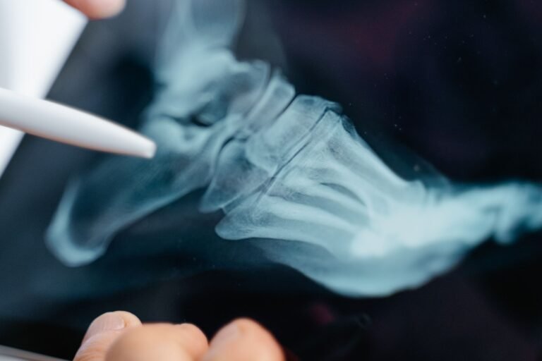
Standard X‑rays are the first line and can show most fractures clearly. In complex cases, a CT scan provides a 3‑dimensional view, while MRI is useful for detecting hairline fractures and soft‑tissue injuries.
Bone scans are rarely needed but can help locate stress fractures that are not visible on X‑ray.
Treatment Options
Treatment depends on the type, location, and severity of the fracture, as well as the patient’s overall health.
Non‑Surgical Management
Most simple fractures heal well with immobilization. Common methods include:
- Cast – A hard plaster or fiberglass shell that holds the bone in place.
- Splint – A removable support used initially or for less severe breaks.
- Brace – Flexible support for fractures near joints, allowing limited movement.
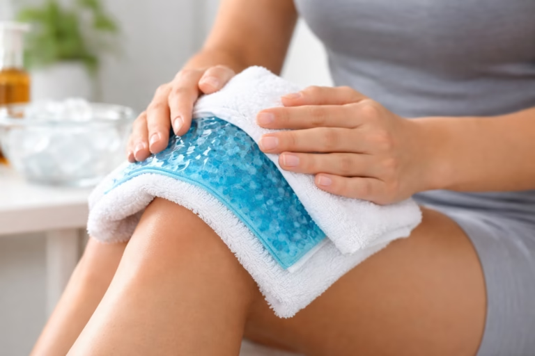
Patients are usually advised to keep the affected limb elevated, apply ice for swelling, and take over‑the‑counter pain relievers as directed.
Surgical Intervention
Complex fractures may need surgery to realign and stabilize the bone. Techniques include:
- Internal fixation – Screws, plates, or rods placed inside the body.
- External fixation – A frame outside the limb connected to pins that go through the skin into the bone.
- Intramedullary nailing – A metal rod inserted into the marrow canal of long bones.
Post‑surgery, a cast or brace may still be used, and physical therapy begins once the surgeon clears the patient.
Recovery and Rehabilitation
Healing time varies widely. A simple wrist fracture may take 6‑8 weeks, while a severe femur break can require 4‑6 months of rehab.
Key components of a successful recovery include:
- Follow‑up appointments – X‑rays to ensure the bone is healing correctly.
- Gradual weight‑bearing – Starting with light activities and increasing as tolerated.
- Physical therapy – Targeted exercises to restore strength, flexibility, and balance.
- Nutrition – Adequate calcium, vitamin D, and protein support bone repair.
- Safety – Using assistive devices like crutches or walkers until confidence returns.
Patients should avoid smoking and limit alcohol, as both can slow bone healing.
Bone Healing Process
Cellular Phases
The body initiates fracture repair through a well‑defined sequence of cellular events. Immediately after injury, blood vessels rupture and a hematoma forms, providing a scaffold for inflammatory cells. Within days, macrophages and neutrophils clear debris while releasing cytokines that attract fibroblasts and stem cells. These cells differentiate into chondrocytes and osteoblasts, laying down a soft callus of cartilage that bridges the fracture gap.
Factors Influencing Healing
Several intrinsic and extrinsic factors can accelerate or impede the healing cascade. Adequate nutrition, particularly sufficient protein, calcium, vitamin D, and vitamin C, supplies the raw materials for new bone formation. Systemic conditions such as diabetes, smoking, and chronic steroid use impair vascular supply and cellular function, often resulting in delayed union. Mechanical stability is also critical; excessive motion disrupts callus formation, while appropriate fixation promotes orderly remodeling.
- Optimized nutrition: balanced diet rich in bone‑supporting nutrients
- Control of chronic diseases: tight glycemic control for diabetics
- Avoidance of tobacco and excessive alcohol
Monitoring Progress
Clinicians track healing through serial imaging and functional assessments. Plain radiographs taken at two‑week intervals can reveal transition from soft to hard callus, indicating progression toward consolidation. Advanced modalities such as CT or MRI are reserved for complex cases where subtle nonunion or malalignment is suspected. Physical examination focuses on pain levels, weight‑bearing tolerance, and range of motion to gauge functional recovery.

Complications of Fractures
Nonunion and Delayed Union
When the normal healing timeline is disrupted, a fracture may fail to unite (nonunion) or take longer than expected (delayed union). Risk factors include severe soft‑tissue injury, inadequate stabilization, and poor vascular supply. Clinically, patients report persistent pain at the fracture site and may exhibit palpable movement at the fracture line.
Infection
Open fractures, where bone is exposed to the external environment, carry a high risk of bacterial contamination. Early administration of broad‑spectrum antibiotics, thorough debridement, and timely wound closure are essential to prevent osteomyelitis. Signs of infection include increasing redness, swelling, warmth, drainage, and systemic symptoms such as fever.
- Open fracture management: immediate irrigation, debridement, and antibiotics
- Closed fracture vigilance: monitor for fever and wound changes
- Long‑term follow‑up: repeat imaging if healing stalls
Compartment Syndrome
Elevated pressure within a closed muscle compartment can compromise circulation and nerve function, representing a surgical emergency. It most often follows high‑energy tibial fractures or severe crush injuries. Early symptoms include disproportionate pain on passive stretch, paresthesia, and a tense, shiny compartment appearance.
Special Considerations in Specific Populations
Children and Adolescents
Growing bones possess a unique structure called the growth plate, which is vulnerable to injury. Fractures that involve the physis require careful alignment to prevent growth disturbances or angular deformities. Pediatric bones remodel more rapidly than adult bones, allowing for slightly greater tolerance of residual angulation.
- Assess physeal involvement: use Salter‑Harris classification
- Prefer minimally invasive fixation when possible
- Schedule regular follow‑up until skeletal maturity
Elderly Patients
Age‑related bone loss, particularly osteoporosis, predisposes seniors to low‑energy fractures such as hip or vertebral compression injuries. Management emphasizes both fracture stabilization and strategies to improve bone density, including pharmacologic agents like bisphosphonates and weight‑bearing exercise programs. Fall‑prevention measures, such as home safety assessments and vision correction, are integral to reducing repeat injuries.
Athletes
High‑performance individuals often sustain fractures from sport‑specific forces, and rapid return to activity is a priority. Surgical fixation may be favored to allow earlier mobilization, but the decision balances the risk of hardware irritation against the need for stability. Rehabilitation protocols are tailored to the sport, incorporating sport‑specific drills, proprioceptive training, and gradual load progression to restore confidence and performance levels.
- Early functional bracing when appropriate
- Gradual re‑introduction of sport‑specific movements
- Psychological support to address fear of re‑injury
Frequently Asked Questions
Can a fracture heal without a cast?
Some minor hairline fractures may heal with rest and protective splinting, but most require some form of immobilization to keep the bone aligned.
How long does a typical fracture take to heal?
Healing time depends on the bone involved, the patient’s age, and overall health. Most fractures in adults heal in 6‑12 weeks, while children often heal faster.
Is it safe to exercise with a cast?
Light, low‑impact activities may be allowed with a doctor’s approval. High‑impact or weight‑bearing exercises should be avoided until the cast is removed and the bone is stable.
What signs indicate a complication?
Increasing pain, swelling, redness, fever, or a change in skin color can suggest infection, especially with an open fracture. Seek medical attention immediately if these occur.
The Bottom Line
Fractures are breaks in bone that range from tiny cracks to severe, open injuries. Prompt recognition, proper medical evaluation, and appropriate treatment, whether casting or surgery, are essential for optimal healing. Supporting recovery with nutrition, safe movement, and follow‑up care helps return you to daily activities with confidence.
Was this page helpful?
Thanks for your feedback! What could we improve?
Thank you! Your feedback helps us improve our articles.
True Impact Health articles are written by our editorial team and based on reliable sources such as public health agencies, clinical guidelines and peer-reviewed research. This article has not yet been medically reviewed. Read our editorial process.
This content is for general information and is not a substitute for professional medical advice, diagnosis or treatment.