Introduction
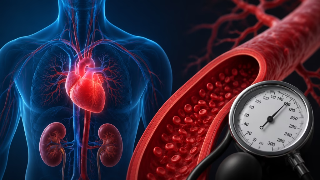
Congestive heart failure (CHF) is a serious cardiovascular condition that affects millions of people worldwide. Despite its name, heart failure does not mean that the heart has completely stopped working. Instead, it means that the heart is unable to pump blood effectively enough to meet the body’s demands. As a result, blood and fluid can accumulate in various parts of the body, including the lungs, legs, ankles, and abdomen.
- Echocardiogram is essential for diagnosing congestive heart failure by assessing heart structure and function in real time.
- Ejection fraction measurement identifies systolic dysfunction; EF below 40% often indicates heart failure with reduced ejection fraction.
- Echo detects ventricular enlargement, wall thickening, diastolic dysfunction, valve disease, and estimates pulmonary artery pressure to evaluate causes and severity.
- Noninvasive, radiation free, and widely available for monitoring, but image quality and operator skill can limit diagnostic accuracy.
Heart failure is a progressive condition that can significantly impact quality of life if left untreated. Common symptoms such as shortness of breath, persistent fatigue, swelling, and exercise intolerance can interfere with daily activities and become increasingly severe over time.
Fortunately, advances in medical imaging have made it possible to diagnose heart failure earlier and more accurately than ever before. One of the most important diagnostic tools available today is the echocardiogram, commonly known as an echo. This noninvasive test provides detailed images of the heart’s structure and function, allowing healthcare providers to identify abnormalities that may indicate congestive heart failure.
In this article, we will explore how an echocardiogram works, what information it provides, how it helps diagnose congestive heart failure, its advantages and limitations, and what patients can expect during the testing process.
Understanding Congestive Heart Failure
What Is Congestive Heart Failure?
Congestive heart failure occurs when the heart muscle becomes too weak or too stiff to pump blood efficiently throughout the body. Because blood circulation becomes less effective, fluid may begin to accumulate in tissues and organs.
The term “congestive” refers to this buildup of fluid, which often causes congestion in the lungs and other parts of the body.
Related Posts:


Heart failure can affect one side of the heart or both sides and may develop gradually over months or years. In some cases, it can occur suddenly following a heart attack or severe cardiac event.
How the Heart Normally Functions
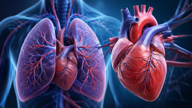
The heart consists of four chambers that work together to circulate blood.
The right side receives oxygen-poor blood from the body and pumps it to the lungs for oxygenation. The left side receives oxygen-rich blood from the lungs and pumps it throughout the body.
When the heart is healthy, this process occurs efficiently and continuously. In heart failure, however, the heart’s pumping or filling ability becomes impaired.
Common Causes of Congestive Heart Failure
Several medical conditions can weaken or damage the heart over time.
Coronary Artery Disease
Coronary artery disease is the most common cause of heart failure. It occurs when plaque buildup narrows the arteries supplying blood to the heart muscle.
Reduced blood flow deprives the heart of oxygen and nutrients, eventually weakening its ability to pump effectively.
High Blood Pressure
Chronic hypertension forces the heart to work harder than normal. Over time, this extra workload causes the heart muscle to thicken and eventually weaken.
Cardiomyopathy
Cardiomyopathy refers to diseases that directly affect the heart muscle. These conditions may result from genetic factors, infections, alcohol abuse, or certain medications.
Heart Valve Disorders
Heart valves regulate blood flow through the heart. Damaged or malfunctioning valves force the heart to work harder and can contribute to heart failure.
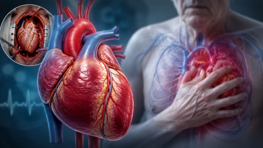
Heart Attacks
A heart attack damages portions of the heart muscle, reducing its ability to contract efficiently and pump blood throughout the body.
Symptoms of Congestive Heart Failure
Recognizing the symptoms of heart failure is important because early diagnosis often leads to better outcomes.
Shortness of Breath
Many patients first notice difficulty breathing during physical activity. As heart failure progresses, shortness of breath may occur even while resting or lying down.
This symptom develops because fluid accumulates within the lungs, making oxygen exchange more difficult.
Persistent Cough or Wheezing
Fluid buildup in the lungs can produce a chronic cough or wheezing. Some patients may cough up white or pink frothy mucus.
Swelling of the Legs, Feet, and Abdomen
When the heart cannot pump effectively, fluid may accumulate in tissues. Swelling often develops in the ankles, feet, legs, and abdomen.
Fatigue and Weakness
Reduced blood flow means muscles and organs receive less oxygen. As a result, patients often experience persistent fatigue and decreased physical endurance.
Rapid or Irregular Heartbeat
The heart may attempt to compensate for reduced pumping efficiency by beating faster. This can lead to palpitations or abnormal heart rhythms.
Why Early Detection Matters
Early diagnosis of congestive heart failure offers several important benefits.
Improved Treatment Outcomes
Identifying heart failure before severe damage occurs allows healthcare providers to initiate treatments that slow disease progression.
Reduced Hospitalizations
Proper management can reduce episodes of fluid overload and prevent emergency hospital admissions.
Better Quality of Life
Patients who receive early treatment often experience improved energy levels, reduced symptoms, and greater ability to participate in daily activities.
Prevention of Complications
Timely intervention helps reduce the risk of complications such as kidney dysfunction, arrhythmias, and advanced heart failure.
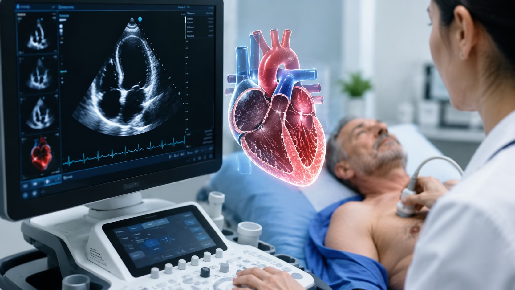
What Is an Echocardiogram?
An echocardiogram is a diagnostic imaging test that uses ultrasound technology to create real-time images of the heart.
Unlike X-rays or CT scans, echocardiography does not use radiation. Instead, it relies on high-frequency sound waves that bounce off cardiac structures and produce detailed images.
A handheld device called a transducer is placed on the chest. The transducer emits sound waves and receives the returning echoes, which are converted into images by a computer.
These images allow doctors to observe the heart as it beats, pumps blood, and moves through its normal cycles.
Types of Echocardiograms
Transthoracic Echocardiogram (TTE)
This is the most commonly performed echocardiogram.
The transducer is placed directly on the chest wall, making the procedure painless and noninvasive.
Most patients undergoing heart failure evaluation receive this type of echocardiogram.
Transesophageal Echocardiogram (TEE)
A transesophageal echocardiogram involves placing a specialized probe into the esophagus.
Because the esophagus lies directly behind the heart, this technique provides exceptionally detailed images.
TEE is often used when standard images are unclear or when evaluating specific cardiac abnormalities.
Doppler Echocardiography
Doppler technology measures the speed and direction of blood flow within the heart.
This information helps detect valve abnormalities and assess blood circulation.
Stress Echocardiogram
A stress echocardiogram evaluates heart function during physical exercise or medication-induced stress.
It helps identify problems that may only become apparent when the heart works harder.
Can an Echocardiogram Detect Congestive Heart Failure?
The simple answer is yes.
An echocardiogram is one of the most important tests used to diagnose congestive heart failure because it provides direct information about how well the heart is functioning.
Measuring Ejection Fraction
One of the most important measurements obtained from an echocardiogram is the ejection fraction.
Ejection fraction refers to the percentage of blood pumped out of the left ventricle during each heartbeat.
A healthy ejection fraction typically ranges between 55% and 70%.
An ejection fraction below 40% often indicates systolic heart failure, meaning the heart muscle has become too weak to pump effectively.
Patients with reduced ejection fraction are often diagnosed with heart failure with reduced ejection fraction (HFrEF).
Assessing Ventricular Size
An echocardiogram can reveal whether the heart chambers have become enlarged.
Enlarged ventricles often indicate chronic pressure overload or cardiomyopathy.
Chamber enlargement is a common finding in many forms of heart failure.
Evaluating Heart Wall Thickness
The test can determine whether the heart muscle walls are abnormally thick.
Thickened walls may develop due to long-standing hypertension or other cardiac conditions.
These changes can impair the heart’s ability to fill properly.
Detecting Diastolic Dysfunction
Not all heart failure occurs because the heart is weak.
Some patients have normal pumping strength but impaired relaxation of the heart muscle.
This condition is known as diastolic heart failure or heart failure with preserved ejection fraction (HFpEF).
An echocardiogram can evaluate how effectively the heart relaxes and fills between contractions.
Identifying Valve Problems
Heart valve disease can contribute significantly to heart failure.
The echocardiogram can identify:
- Valve narrowing (stenosis)
- Valve leakage (regurgitation)
- Structural valve abnormalities
These findings help determine whether valve disease is contributing to symptoms.
Measuring Pulmonary Artery Pressure
Advanced heart failure often causes increased pressure within the pulmonary circulation.
An echocardiogram can estimate pulmonary artery pressure and identify strain on the right side of the heart.
Signs of Fluid Overload Seen on Echocardiography
Echocardiography may reveal several indicators of fluid accumulation.
Left Atrial Enlargement
An enlarged left atrium often reflects chronically elevated filling pressures within the heart.
Inferior Vena Cava Enlargement
A dilated inferior vena cava may indicate elevated pressure within the venous system and fluid overload.
Pulmonary Congestion Indicators
Certain ultrasound findings may suggest excess fluid within the lungs, supporting a diagnosis of congestive heart failure.
Advantages of Echocardiography
Noninvasive Procedure
No surgery, needles, or incisions are required.
No Radiation Exposure
The procedure is safe and can be repeated as often as necessary.
Real-Time Imaging
Doctors can observe heart function as it occurs.
Widely Available
Most hospitals and cardiology clinics offer echocardiography services.
Useful for Monitoring Progress
Repeated echocardiograms help track disease progression and treatment effectiveness.
Limitations of Echocardiography
Despite its benefits, echocardiography has limitations.
Image Quality Challenges
Obesity, lung disease, and chest wall abnormalities may affect image quality.
Operator Dependency
Results depend on the skill of the sonographer performing the test and the physician interpreting the images.
Early Disease Detection
Very subtle abnormalities may occasionally require more advanced imaging techniques.
Additional Tests Used Alongside Echocardiography
Doctors often combine echocardiography with other diagnostic tests.
Electrocardiogram (ECG)
Evaluates heart rhythm and electrical activity.
Chest X-Ray
Assesses heart size and detects fluid accumulation in the lungs.
Blood Tests
B-type natriuretic peptide (BNP) levels often increase in heart failure.
Cardiac MRI
Provides highly detailed images when additional information is needed.
Cardiac CT Scan
Useful for evaluating coronary arteries and structural abnormalities.
Treatment Following Diagnosis
Treatment depends on the severity and underlying cause of heart failure.
Common therapies include:
Medications
- ACE inhibitors
- ARBs
- Beta-blockers
- Diuretics
- Mineralocorticoid receptor antagonists
- SGLT2 inhibitors
Lifestyle Changes
- Low-sodium diet
- Fluid management
- Regular physical activity
- Weight control
- Smoking cessation
Advanced Therapies
Some patients may require:
- Implantable cardioverter-defibrillators (ICDs)
- Cardiac resynchronization therapy
- Ventricular assist devices
- Heart transplantation
Conclusion
An echocardiogram is one of the most valuable tools available for diagnosing congestive heart failure. By providing detailed information about heart structure, pumping ability, chamber size, valve function, and fluid status, it allows healthcare providers to identify heart failure accurately and determine its underlying cause.
Whether evaluating symptoms such as shortness of breath and fatigue or monitoring the progression of established heart failure, echocardiography plays a central role in modern cardiovascular care. Its noninvasive nature, safety profile, and ability to provide real-time information make it indispensable for both diagnosis and long-term management.
If you are experiencing symptoms that may suggest congestive heart failure, speaking with your healthcare provider about an echocardiogram could be an important step toward obtaining an accurate diagnosis and starting effective treatment. Early detection remains one of the most powerful ways to protect heart health and improve long-term outcomes.